Introduction
COHAT stands for comprehensive oral health assessment and treatment. AAHA uses the term, along with COPAT (comprehensive oral prevention, assessment, and treatment), as a more accurate name than "dental" or "prophy" for the procedure done under general anesthesia to prevent and treat periodontitis.1 The credentialed veterinary technician runs much of a COHAT: anesthesia and airway care, positioning, scaling, polishing, sulcal lavage, probing and charting, radiographs and recovery. The veterinarian examines, diagnoses and decides on extractions or other surgery.
This guide walks through the steps in order, names the instrument for each step and the mistake that most often goes with it, and covers patient safety, operator protection and the facts the VTNE likes to test.
Key facts
- General anesthesia with endotracheal intubation, monitoring and physiologic support is necessary for dental cleaning. AAHA calls anesthesia-free dentistry unacceptable.1
- Use a cuffed endotracheal tube of the correct size plus a gauze pharyngeal (throat) pack, and remove the pack before extubation.3
- Do not overinflate the cuff, especially in cats, and disconnect the tube from the breathing circuit before turning the patient.2
- Keep an ultrasonic scaler tip moving and in contact with one tooth for no more than 15 seconds, with water cooling.2
- Polish with a low-speed handpiece and prophy cup at no more than 3000 rpm, with fine paste, and no more than 5 seconds on one tooth at a time.12
- Probe every tooth in at least six places. Probing depth should be no more than 2 to 3 mm in a midsized dog and 1 mm in a midsized cat.1
- Rinse the mouth with 0.12% chlorhexidine before scaling to reduce bacterial aerosols, and wear mask, eye protection and gloves.12
How the VTNE tests this
- Put the steps in order: which comes first, polishing or scaling, lavage or polishing. Domain 3 Task C (perform dental procedures); K21 (dental procedures, including cleaning, charting and preventive procedures).7
- Choose the instrument or setting: curette versus sickle scaler, polishing speed, scaler contact time. K22 (dental equipment, instruments and supplies).7
- Spot the safety problem: deflated cuff, no throat pack, spring gag in a cat, missing PPE, cold patient. K23 (safety considerations related to dental procedures).7
- Read a probing value as normal or abnormal for a dog or a cat, and know what to chart. K21.7
Core concepts
Why anesthesia and the airway come first
A real cleaning has to reach below the gum line. Subgingival scaling is the most important step of the cleaning, because removing plaque above the gum line alone does not treat periodontal disease.2 Probing is a noxious stimulus, radiographs need a motionless patient, and an abrupt response to stimulation risks bite injury, so the work needs general anesthesia.1 A dental procedure also puts a lot of water and debris into the mouth. Airway protection prevents aspiration pneumonia from inhaled cooling water and flushed debris.3
Plaque, calculus and roughened enamel
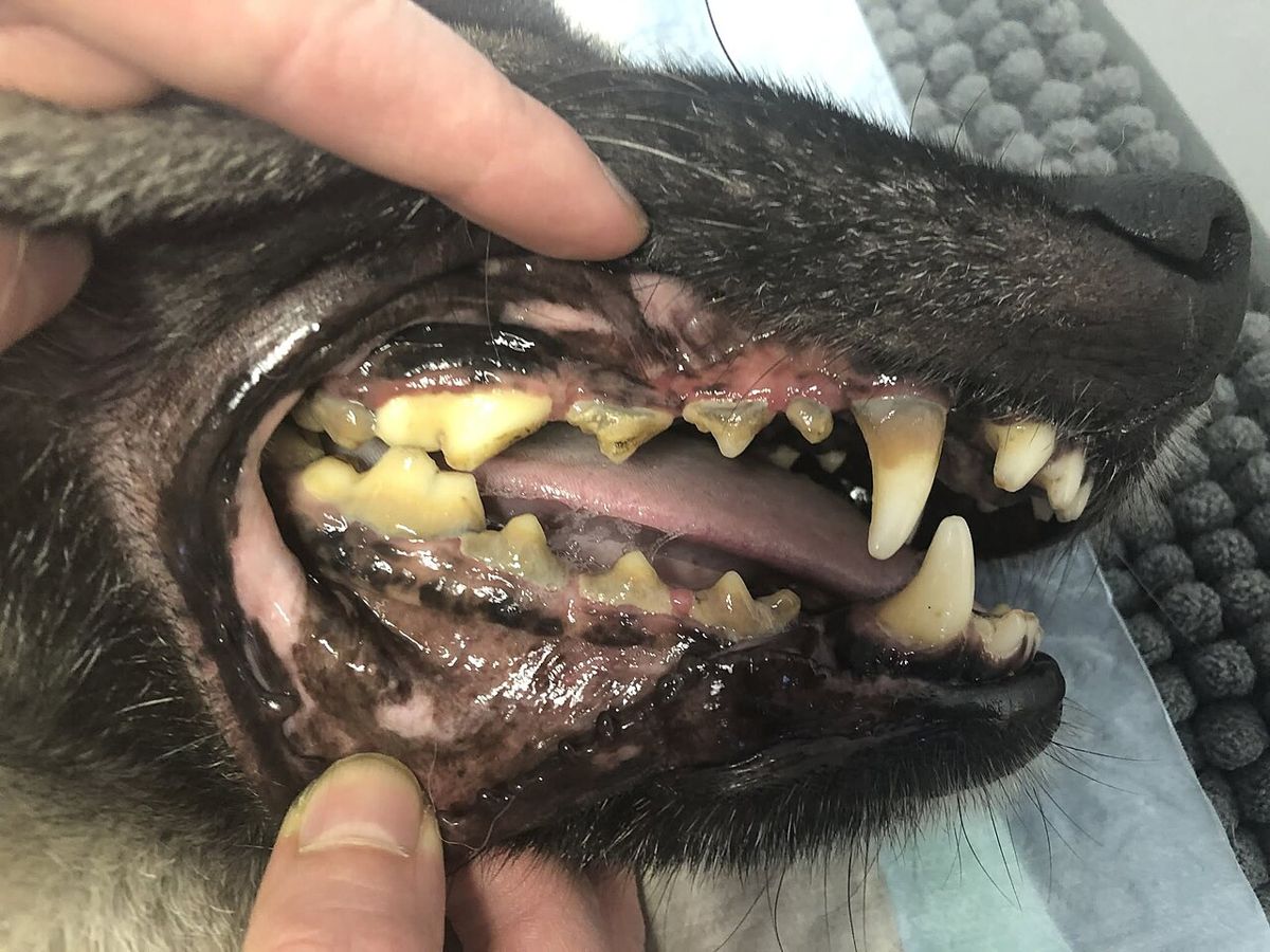
Plaque is the soft bacterial film; calculus is the hardened deposit (Figure 1). Scaling with hand or power instruments leaves microabrasions that roughen the tooth and help plaque stick again. Polishing smooths the surface and slows plaque attachment.2 Scaling and polishing also push calculus and bacteria-laden paste into the sulcus, which is why the sulcus is flushed afterward.2
Hand instruments in one paragraph
A scaler (sickle scaler) is triangular with two cutting edges and a sharp tip. It is for supragingival use only, because its shape, sharp back and tip can easily damage the gingiva.2 A curette has a smooth, rounded heel and toe, so it is less traumatic to soft tissue and is the instrument for subgingival plaque and calculus.1 Hand instruments are held in a modified pen grasp and only work when sharp, which means sharpening at least weekly if they are used regularly.2
Procedure or technique
AAHA and WSAVA list the same core steps, though their order differs a little around probing. AAHA lists probing after scaling and full-mouth radiographs; WSAVA lists sulcal lavage, then probing and charting, then radiographs.12 Follow your hospital protocol. The sequence below is the one most exam questions expect: assess, scale, polish, flush, probe and chart, image, then treat.
- Conscious oral exam and consent. The veterinarian examines the awake patient and shares findings, a plan, fees and consent with the owner before any extra treatment.1 Never extract a tooth without prior owner consent.2
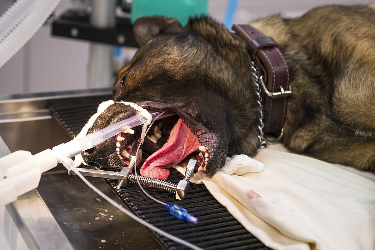
- Anesthesia and airway. Preoxygenate for 2 to 5 minutes before induction.1 After induction, take a quick look at the oropharynx, then intubate and place the throat pack.2 Inflate the cuff to a seal without overinflating it.2 In cats, apply eye lubricant at the start and reapply it during the procedure and recovery.3
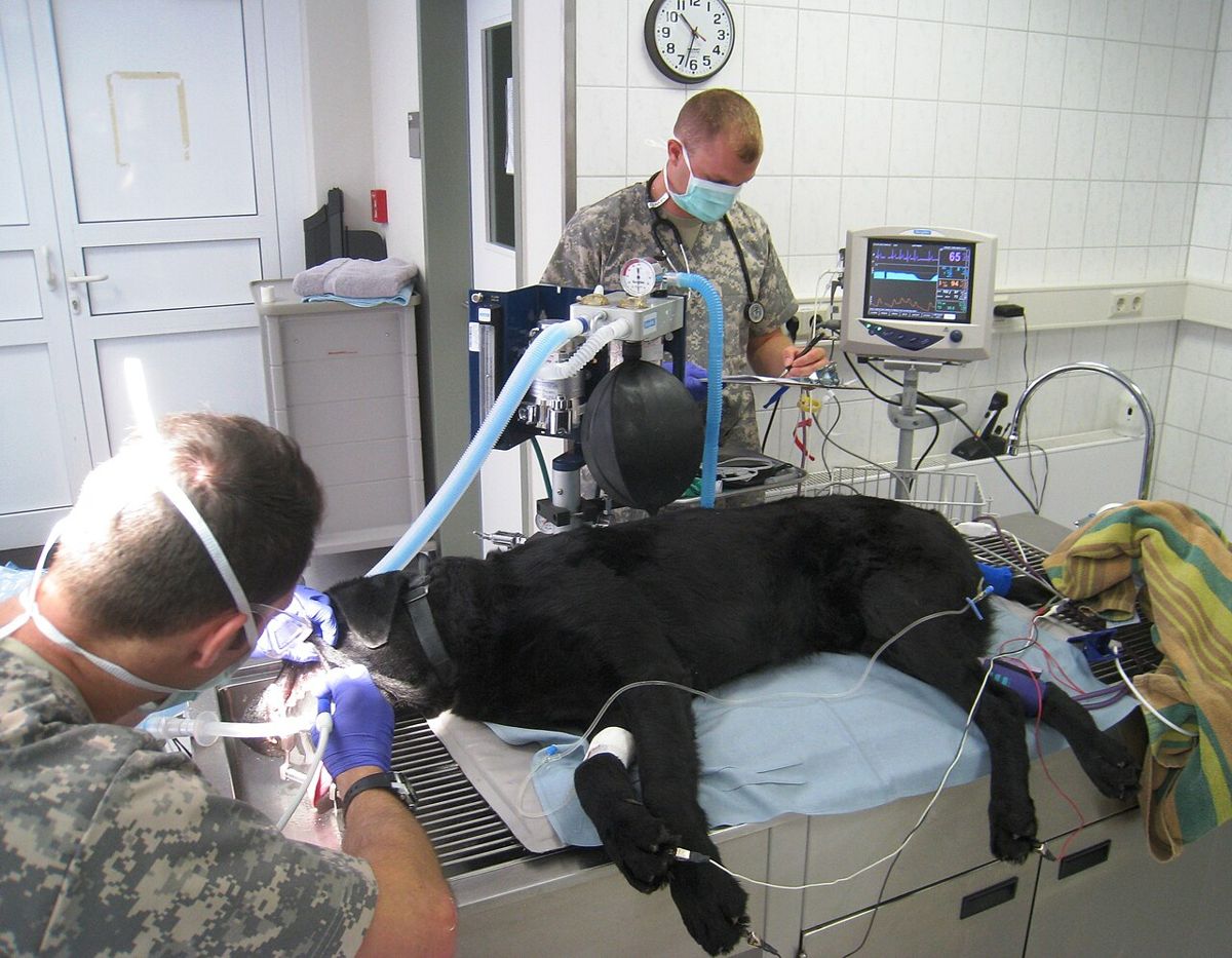
- Position and warm the patient. In lateral recumbency, a towel or sandbag under the neck tilts the head down so saliva, blood and irrigating fluid drain out of the mouth.5 Disconnect the tube from the circuit every time you turn the patient, then recheck the airway and cuff.23 Start active warming (Figure 2 shows a patient positioned for a dental cleaning).
- Photograph and rinse. WSAVA recommends preoperative photos of each side and the front. Rinse the mouth with 0.12% chlorhexidine to lower the bacterial load.2
- Supragingival scaling. Use an ultrasonic scaler with water spray, or a hand scaler above the gum line. Large deposits can be removed carefully with calculus forceps.2 Use a light touch with the side of the tip, keep it moving in overlapping sweeps, and stay no more than 15 seconds on one tooth.2
- Subgingival scaling. Use a curette below the gum line, or an ultrasonic scaler fitted with a low-powered periodontal tip followed by a curette.12 Standard tips must not go under the gingival margin, because the water does not reach the working end and the tooth can overheat.2 AAHA says not to use a rotary scaler, which roughens the tooth too much.1
- Check for residual calculus. Run an explorer over the teeth feeling for rough spots, or dry the teeth with air: leftover calculus looks chalky. A disclosing solution also works.2
- Polish. Low-speed handpiece, prophy angle and rubber cup at no more than 3000 rpm, with prophy paste or fine pumice. Medium or coarse grit causes enamel loss.1 Always use enough paste, since a dry cup overheats the tooth, and press lightly so the cup edge flares into the sulcus.2 Use disposable prophy angles and single-patient paste to avoid cross contamination.1
- Sulcal lavage (irrigation). Gently flush each sulcus with a blunt 22 to 25 gauge cannula. Sterile saline works; most dentists prefer 0.12% chlorhexidine.2 Then use the air or water syringe to look for remaining calculus under the gum.1
- Probe and chart. Start at the first incisor of one quadrant and move distally one tooth at a time so no tooth is skipped. Walk the probe gently around the tooth and measure six sites per tooth.2 Charting goes faster four-handed, with one person probing and calling out findings and one recording.2
- Full-mouth intraoral radiographs with film or a digital system.1
- Treatment by the veterinarian: periodontal therapy, extractions, biopsy of abnormal masses. AAHA lists barrier sealants as an option to reduce plaque build-up; they do not replace home care or future cleanings.1
- Recovery. Remove the throat pack before extubation.3 Keep the patient intubated until it swallows and is sternal, keep it warm and keep monitoring until it is awake.1 AAHA's anesthesia guideline suggests the nose slightly lower than the back of the head and the cuff left slightly inflated at extubation, to pull blood clots and debris out of the trachea.4
- Home care teaching at discharge, pointing owners to VOHC-accepted products.1
| Step | Instrument or supply | Common error |
|---|---|---|
| Airway | Cuffed endotracheal tube, gauze throat pack | Cuff overinflated, or pack left in at extubation |
| Rinse | 0.12% chlorhexidine | Skipped, so more bacteria in the aerosol |
| Supragingival scaling | Ultrasonic scaler, sickle scaler | Tip held still or over 15 seconds on one tooth; sickle scaler used under the gum |
| Subgingival scaling | Curette, or periodontal ultrasonic tip then curette | Standard ultrasonic tip pushed under the gingiva |
| Residual check | Explorer, air syringe | Calculus left behind because the teeth "look clean" |
| Polishing | Low-speed handpiece, prophy angle, cup, fine paste | Over 3000 rpm, coarse paste, dry cup, or skipped |
| Sulcal lavage | Blunt 22 to 25 gauge cannula, saline or chlorhexidine | Skipped, leaving paste and debris in the sulcus |
| Probing and charting | Periodontal probe, dental chart | Fewer than six sites, or teeth skipped |
Source: AAHA 2019 Dental Care Guidelines; WSAVA Global Dental Guidelines; 2025 FelineVMA oral health guidelines.123
Flowchart
Species differences
- Normal probing depth: up to 2 to 3 mm in a midsized dog and 1 mm in a midsized cat (AAHA). WSAVA gives 0 to 3 mm in dogs and 0 to 0.5 mm in cats.12 A depth that is normal in a dog can be a pocket in a cat.
- Cuff care in cats: overinflation can cause tracheal damage or necrosis, especially when the patient is moved.2 Repositioning raises the risk of tracheal tears, so disconnect before turning and recheck the cuff after.3
- Mouth gags in cats: spring-loaded gags can compress the maxillary artery and have caused blindness and other neurological deficits. Avoid them, or use a prop that does not hyperextend the jaws.3
- Eyes and heat in cats: their large eyes need lubricant throughout. Water around the head can cause rapid hypothermia, so active warming and temperature checks are needed.3
- Timing: AAHA recommends the first complete cleaning with radiographs by 1 year of age for cats and small to medium breed dogs, and by 2 years for larger breeds.1
Calculation
How many probing readings go on the chart? An adult dog has 42 permanent teeth, and each tooth is probed at six sites.2
42 teeth x 6 sites per tooth = 252 sites
The "teeth" units cancel and the answer is in sites. An adult cat with all 30 teeth: 30 teeth x 6 sites per tooth = 180 sites. Numbers like these are why four-handed charting saves time.
Safety, legal and client education
| Hazard | Protection |
|---|---|
| Bacterial aerosol from scaling and polishing | Chlorhexidine rinse first; mask, eye protection and gloves for the operator and anyone close by13 |
| Calculus and debris flying toward the eyes | Protective spectacles or face shield whenever a drill or scaler is used5 |
| Contaminated dental unit water | Bacterial water filter or chlorhexidine flushing of the system2 |
| Radiation from the primary beam, scatter and tube head leakage | Lead apron, thyroid shield and dosimeter1 |
| Waste anesthetic gas | Scavenging system and a properly placed endotracheal tube1 |
| Chronic strain injury | Proper seating, fatigue mats, supplies at working height, swivel stool1 |
Source: AAHA 2019 Dental Care Guidelines; WSAVA Global Dental Guidelines; FelineVMA 2025; RVC dentistry workplace notes.1235
- Room choice: dental work belongs in its own space, not in the sterile surgical suite and not near sick patients or clean procedures.12
- Scope of practice: AAHA notes that assistants should only do tasks that fit their skill level and the state practice act.1 Extractions and diagnosis belong to the veterinarian.
- Client talk about anesthesia-free cleaning: cleaning only the visible crowns gives the owner a false sense that the pet's mouth is healthy.1
- Toothpaste: human toothpastes are not recommended for pets because detergents or fluoride can cause stomach upset or fluorosis if swallowed.2 Professional fluoride treatment is one option a veterinarian may use for enamel defects.6
Common mistakes and exam traps
- "The teeth look clean, so skip polishing." Scaling roughens enamel. Skipping polishing leaves a surface that collects plaque faster.2
- Faster is not better. Polishing above 3000 rpm, or with a dry cup, overheats the tooth and does not clean any better.2
- Sickle scaler under the gum. The sharp tip and back cut the gingiva; the curette is the subgingival instrument.12
- Using the point of the ultrasonic tip. Use the side, with light pressure, keep it moving and keep the water running.2
- Pack left in. Removing the throat pack before extubation is a safety priority.3 Count it in and out the same way you count sponges.
- Cat probing depth read with dog numbers. 2 mm is normal for a dog but over the AAHA figure of 1 mm for a cat.1
Memory aids
Scale, Smooth, Sweep, Sound (memory aid): scale the calculus, smooth by polishing, sweep the sulcus with lavage, then sound (probe) every tooth. It keeps polishing after scaling and lavage after polishing.
15 and 5 (memory aid): no more than 15 seconds of ultrasonic contact on one tooth, and no more than 5 seconds of polishing on one tooth at a time.
Three thousand, not three times faster (memory aid): the prophy cup runs at no more than 3000 rpm.
Six around, one to three deep (memory aid): six probing sites around each tooth; normal depth up to 1 mm in a cat and up to 2 to 3 mm in a dog.
Pack in, pack out (memory aid): the throat pack goes in after intubation and comes out before extubation.
Quick review
- A COHAT is done under general anesthesia with a cuffed endotracheal tube; anesthesia-free cleaning is not acceptable.
- Check the cuff and place the throat pack before any water enters the mouth.
- Do not overinflate the cuff in cats; disconnect from the circuit before turning the patient.
- Tilt the head down so fluid drains, and keep the patient warm and dry.
- Rinse with 0.12% chlorhexidine before scaling to reduce aerosolized bacteria.
- Ultrasonic scaler: side of the tip, light touch, moving, water on, 15 seconds per tooth maximum.
- Sickle scaler above the gum, curette below the gum.
- Polish at no more than 3000 rpm with fine paste, enough paste, 5 seconds per tooth at a time.
- Flush the sulcus with a blunt cannula after polishing.
- Probe six sites per tooth, starting at the first incisor and moving distally.
- Remove the throat pack before extubation; extubate when the patient swallows and is sternal.
- Wear mask, eye protection and gloves; use lead, thyroid shield and dosimeter for radiographs.