Introduction
Oral pathology means disease of the teeth, gums and mouth. During a dental procedure the credentialed veterinary technician is often the first person to see a cracked crown, a tooth resorption lesion or a mass. The technician explores, probes, takes radiographs and charts what is found. The veterinarian diagnoses and decides which teeth are treated or extracted.
This guide covers the lesions you will see most: tooth fractures, feline tooth resorption, feline chronic gingivostomatitis, malocclusion classes 1 to 4, persistent deciduous teeth, oronasal fistula and oral masses. It then covers how you assist with extractions, the local anesthetic dose math for dental nerve blocks, and what owners need to hear at discharge. Use the standard AVDC (American Veterinary Dental College) names and abbreviations when you chart, because the VTNE uses them too.1
Key facts
- A complicated fracture exposes the pulp. An uncomplicated fracture does not.1 In a mature tooth, every fracture that exposes the pulp needs root canal therapy or extraction.4
- More than 60% of cats will have tooth resorption at some point in their lives. The mandibular third premolars (307 and 407) are the teeth affected most often.2
- Tooth resorption has 5 stages (how much tooth is lost) and 3 radiographic types (what happens to the periodontal ligament space).1
- In feline chronic gingivostomatitis, the key sign is inflammation of the caudal oral mucosa, at or lateral to the palatoglossal folds. Partial-mouth or full-mouth extraction is the only treatment that gives lasting improvement.3
- Class 1 malocclusion means the jaws are normal and one or more teeth are out of position. Classes 2 and 3 are skeletal: the lower jaw sits too far back (class 2) or too far forward (class 3). Class 4 is an asymmetric skeletal malocclusion.1
- A persistent deciduous tooth is one still present when it should have exfoliated. It should be removed as soon as possible without damaging the permanent tooth.15
- Take a radiograph after every extraction to confirm that no root is left behind.8
How the VTNE tests this
Dentistry is Domain 3 of the 2023 VTNE content outline, with 10 of the 150 scored items (7%). Its tasks include applying anatomy, physiology and pathophysiology to dentistry, preparing dental equipment and supplies, performing the COHAT and producing diagnostic dental radiographs.12 Expect questions like these:
- Name the lesion. A crown fracture with a red dot in the center, a cat with a pink tissue defect at the gumline, a dog whose lower canines hit the palate.
- Pick the class or stage. Malocclusion class, fracture type, or tooth resorption stage and type from a short description.
- Know why radiographs matter. Resorption type, retained roots and periapical lesions can only be seen on a radiograph.
- Assist safely. Instrument order for an extraction, suture choice, nerve block dose limits, and discharge instructions.
Core concepts
Tooth fractures
The AVDC sorts tooth fractures by what tissue is involved and whether the pulp is open.1 The pulp is the living tissue inside the tooth: nerves and blood vessels. An exposed pulp bleeds only briefly. After that it looks like a red dot if the pulp is still alive, or a black hole if it has died.4 An untreated complicated crown fracture leads to a periapical abscess.4 Do not dismiss uncomplicated fractures either. The AVDC cites a study in which nearly 1 in 4 patients with uncomplicated crown fractures had a periapical lesion on radiographs.13
A tooth with no fracture can still be dead. A discolored tooth (pink, purple, gray or green) indicates endodontic disease, and 87% to 92% of these teeth are nonvital.4 Chart every discolored tooth.
| Fracture (abbreviation) | What is involved | Pulp exposed? |
|---|---|---|
| Enamel infraction (T/FX/EI) | Crack in the enamel, no tooth lost | No |
| Enamel fracture (T/FX/EF) | Crown lost, limited to enamel | No |
| Uncomplicated crown fracture (T/FX/UCF) | Crown, into dentin | No |
| Complicated crown fracture (T/FX/CCF) | Crown | Yes |
| Uncomplicated crown-root fracture (T/FX/UCRF) | Crown and root | No |
| Complicated crown-root fracture (T/FX/CCRF) | Crown and root | Yes |
| Root fracture (T/FX/RF) | Root only | Varies |
Source: AVDC Nomenclature.1
Feline tooth resorption
Tooth resorption (TR) is the loss of hard tooth tissue. Older names include FORL (feline odontoclastic resorptive lesion) and cervical line lesion. No true cause has been identified. TR is more common in older and purebred cats.2 Lesions usually start on the root surface and show up at the gumline as inflamed granulation tissue filling a defect, often with a sharp ledge of enamel at the edge.2 Lesions open to the mouth can be painful.2
Stage 1 lesions cannot be seen on radiographs. You find them by running the explorer end of the probe-explorer along the cementoenamel junction (CEJ), where crown enamel meets root cementum. Gingivitis on one tooth in a mouth without periodontitis can be a clue to a lesion under the gum.2
| Grade | What it means |
|---|---|
| Stage 1 | Mild loss of cementum, or cementum and enamel |
| Stage 2 | Moderate loss into dentin, not reaching the pulp cavity |
| Stage 3 | Deep loss into the pulp cavity; most of the tooth is still intact |
| Stage 4 | Extensive loss into the pulp cavity; most of the tooth has lost its integrity (4a, 4b, 4c) |
| Stage 5 | Only irregular radiopaque remnants; gingiva fully covers the site |
| Type 1 | Focal radiolucency with a normal periodontal ligament (PDL) space |
| Type 2 | PDL space narrowed or gone; the root looks like it is being replaced by bone |
| Type 3 | Features of type 1 and type 2 in the same tooth |
Source: AVDC Nomenclature and the Merck Veterinary Manual.12
Type matters because it changes treatment. Most resorbing teeth should be extracted. Crown amputation, which removes the crown and leaves the resorbing root in place on purpose, is an option only for radiographically confirmed type 2 lesions with no periodontitis, endodontic disease or stomatitis.2 That is why the radiograph is not optional.
See figure 2: tooth resorption on tooth 407 in a cat (Joel Mills, CC BY-SA 3.0, Wikimedia Commons).

Feline chronic gingivostomatitis (FCGS)
FCGS (also called feline stomatitis) is a painful, progressive inflammation of the oral mucosa. It is thought to be an overactive immune response to plaque bacteria.3 Feline calicivirus is strongly linked: up to 100% of affected cats in some studies are chronic carriers.3 What you see: ulcerated, proliferative tissue in the caudal mouth, at and lateral to the palatoglossal folds, plus halitosis, drooling and difficulty eating. A hungry cat may approach the food bowl, then hiss and run away. Many cats are too painful to examine awake.3 If the inflammation is confined to the gingiva around the teeth, think gingivitis or periodontitis instead.3
Treatment removes the plaque-retentive surfaces, meaning the teeth. Partial-mouth extraction (all premolars and molars) or full-mouth extraction is the only treatment that gives lasting control. When full-mouth extraction is done early and postoperative radiographs confirm no retained roots, 60% to 80% of cats appear cured or greatly improved.3 A retained root fragment can prevent improvement, so full-mouth radiographs are essential.3 Atypical lesions, such as a one-sided proliferative mass, need a biopsy to rule out cancer.3
See figure 3: chronic gingivostomatitis in a cat (Anja Bacher-Bröhling and Uwe Gille, CC BY-SA 4.0, Wikimedia Commons).

Malocclusion
Occlusion is how the teeth meet when the mouth closes. Treat a malocclusion when abnormal contact is injuring soft tissue or other teeth.9
- Class 1 (MAL1), neutroclusion: normal jaw relationship, but one or more teeth are malpositioned.1 Example: linguoversion of the mandibular canines (tipped toward the tongue), which can injure the palate.9 A rostral crossbite, with a lower incisor sitting in front of the upper incisors, is also class 1.1
- Class 2 (MAL2), mandibular distoclusion: the lower arch sits caudal to normal (overbite). The lower canines often traumatize the palate.19
- Class 3 (MAL3), mandibular mesioclusion: the lower arch sits rostral to normal (underbite).1
- Class 4 (MAL4), maxillomandibular asymmetry: asymmetry front to back, side to side, or up and down. Up and down asymmetry gives an open bite.1
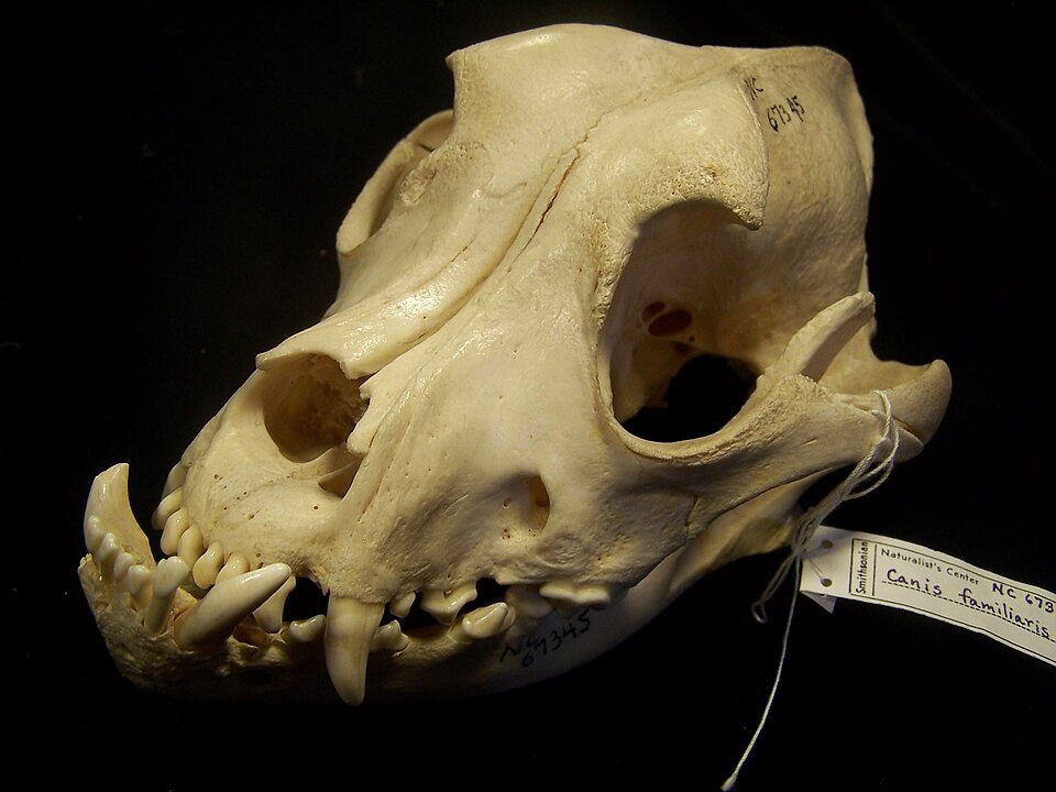
Persistent deciduous teeth
The AVDC prefers "persistent deciduous tooth" over "retained deciduous tooth" because "retained" can be confused with an unerupted tooth.1 Persistent deciduous teeth are common in dogs, especially small breeds such as Toy Poodles. They can push the permanent tooth out of position, causing malocclusion, or trap food and lead to periodontal disease. They should be removed as soon as possible, taking care not to damage the developing permanent tooth.5
Oronasal fistula
An oronasal fistula (ONF) is an acquired opening between the mouth and nasal cavity along the upper dental arch.1 Only a thin plate of bone separates the maxillary canine root from the nasal cavity, so this is a common site.8 Deep palatal pockets from periodontal disease can erode through. These teeth need extraction and fistula repair.7 An ONF is also a possible complication of a poorly performed extraction.8 Owners typically report bouts of sneezing with a bloody nasal discharge. Probe the palatal (medial) side of the maxillary canines carefully.11
Oral masses
"Epulis" is a general term for any gingival mass, not a diagnosis.1 A mass cannot be identified by its look, so biopsy is essential.6
| Lesion | Signs the technician sees | Likely treatment (veterinarian decides) |
|---|---|---|
| Complicated crown fracture | Red dot or black spot in the fractured crown | Root canal therapy or extraction4 |
| Tooth resorption (cat) | Pink tissue in a crown defect at the CEJ; explorer catches | Extraction; crown amputation for type 2 only2 |
| FCGS (cat) | Caudal mouth ulceration, drooling, pain on opening | Partial or full-mouth extraction3 |
| Persistent deciduous tooth | Two teeth of the same type in one spot | Prompt extraction of the deciduous tooth5 |
| Oronasal fistula | Sneezing, bloody nasal discharge, deep palatal pocket on a maxillary canine | Extraction and fistula repair711 |
| Peripheral odontogenic fibroma | Firm gingival mass, usually dogs over 6 years | Excision; complete excision appears curative6 |
| Canine acanthomatous ameloblastoma | Gingival mass invading bone, may move teeth | Surgery; locally aggressive68 |
| Malignant melanoma, SCC, fibrosarcoma | Pigmented or nonpigmented, ulcerated or proliferative mass | Biopsy, then surgery and other cancer treatment6 |
Sources: Merck Veterinary Manual, AVDC, WSAVA, Kirk and Bistner's Handbook.
Procedure or technique: assisting with extractions
Extraction is indicated for many of the lesions above: endodontic disease, severe periodontal disease, traumatic malocclusion, persistent deciduous teeth, tooth resorption and caudal stomatitis.8 The aim is to cut the periodontal ligament, the fibers that hold the root in its socket (alveolus), and remove the whole root.10 Always confirm the client has consented to extractions.10
- Analgesia and radiographs. Give preoperative analgesia, perform regional nerve blocks, and take radiographs before every extraction. They show curved roots, resorption, ankylosis and extra roots.10
- Incise the attachment. The gingival attachment is cut 360 degrees around the tooth with a No. 15 scalpel blade.10
- Section multirooted teeth. Two-rooted teeth are usually cut in half so each root comes out on its own.8
- Elevate. The elevator goes between tooth and alveolar bone and is held with slow, steady pressure to fatigue the ligament. Patience prevents fractured roots.10
- Remove. Extraction forceps are used only when the tooth is very loose, rotating along its long axis without excessive force.10
- Radiograph again. A postoperative radiograph documents complete removal. The WSAVA cites a study that found retained roots in 92% of extracted carnassial teeth in dogs and cats.8
- Close. Use 3-0 to 5-0 absorbable monofilament suture on a reverse cutting needle, simple interrupted, 2 to 3 mm apart, with no tension on the flap.8
Regional nerve blocks
The WSAVA describes the inferior alveolar, palatine, infraorbital, middle mental and maxillary blocks. Volumes are small: 0.1 to 0.2 mL in cats and 0.2 to 1 mL in dogs, and the total dose of all blocks combined must stay under the toxic dose.8 In cats and brachycephalic dogs the infraorbital canal is only a few millimeters long, so the needle is advanced only about 2 mm to avoid the eye.8 A mandibular block can numb the rostral two-thirds of the tongue, so watch for tongue chewing in recovery.8 Technique details are in the local and regional anesthesia guide (Domain 9).
Species differences
- Cats: TR and FCGS are mainly feline problems.23 SCC is the most common malignant oral tumor in cats, and melanoma is very rare in the feline mouth.6 Use the smaller block volumes.8
- Dogs: The three most common malignant oral tumors are malignant melanoma, SCC and fibrosarcoma, and they are more common in dogs over 8 years old.6 Persistent deciduous teeth are most common in small breeds.5 Tooth resorption also occurs in dogs, mainly on premolars and molars.2
- Tumor location: The more caudal a malignant tumor sits in the mouth, the poorer the prognosis.8
Calculation
Maximum bupivacaine volume for a cat. The WSAVA suggests a maximum bupivacaine dose of 2 mg/kg in cats and 4 mg/kg in dogs.8 A 4 kg cat needs four dental blocks with 0.5% bupivacaine.
- 0.5% means 0.5 g per 100 mL, which is 5 mg/mL.
- Maximum dose: 4 kg x 2 mg/kg = 8 mg.
- Maximum volume: 8 mg / 5 mg/mL = 1.6 mL total for all blocks.
- Planned: 4 blocks x 0.2 mL = 0.8 mL (4 mg), which is within the limit.
Always add the blocks together. The limit is for the whole patient, not for each site.
Safety, legal and client education
- Diagnosis and extraction are the veterinarian's decisions. The technician records findings and assists. Extraction requires the client's consent.10
- Pain control at home. Patients go home with analgesics, such as an NSAID alone or with an opioid, unless contraindicated.10
- Diet. Offer only soft food and no hard chews or treats for 5 to 7 days after extraction.10
- FCGS owners. Explain that the goal is to remove plaque-retentive surfaces, that most cats improve, and that some need ongoing medication after surgery.3
- When to call. Sneezing or nasal discharge after a maxillary extraction may mean an oronasal fistula.811
Common mistakes and exam traps
- "Uncomplicated" does not mean "ignore." Radiograph it; periapical lesions are common.13
- Stage versus type. Stage is depth of tissue loss (1 to 5). Type is the radiographic PDL pattern (1 to 3).1
- Stage 1 TR is invisible on radiographs. Only the explorer finds it.2
- Class 1 versus class 2. Lower canines hitting the palate can be either. If the jaw lengths are normal it is class 1; if the mandible is short it is class 2.19
- Epulis is not a diagnosis. Only a biopsy tells benign from malignant.16
- Skipping the post-extraction radiograph. Retained roots are common and keep FCGS going.38
Memory aids
C for Complicated, C for Canal: a complicated fracture has the pulp canal open, so it needs root canal therapy or extraction (memory aid).
Red dot alive, black hole dead: an exposed pulp that is still vital looks red; a necrotic pulp looks black (memory aid).
Type 2 = Two choices: only type 2 resorption, with no other oral disease, has the second option of crown amputation instead of extraction (memory aid).
2 Back, 3 Forward: class 2 puts the mandible too far back (overbite), class 3 too far forward (underbite) (memory aid).
Two in one spot, pull the baby: a deciduous and a permanent tooth of the same type side by side means the deciduous tooth should come out promptly (memory aid).
Quick review
- Complicated fractures expose the pulp; in a mature tooth they need root canal therapy or extraction.
- 87% to 92% of discolored teeth are nonvital.
- More than 60% of cats develop tooth resorption; 307 and 407 are affected most often.
- Stage 1 TR is found with the explorer at the CEJ, not on radiographs.
- Crown amputation is for type 2 TR only, with no periodontitis, endodontic disease or stomatitis.
- FCGS shows caudal mucosal inflammation lateral to the palatoglossal folds; extraction helps 60% to 80% of cats.
- MAL1 is dental; MAL2 (mandible back), MAL3 (mandible forward) and MAL4 (asymmetry) are skeletal.
- Persistent deciduous teeth should be removed promptly without harming the permanent tooth.
- Oronasal fistulas form most often at the maxillary canine; signs are sneezing and bloody nasal discharge.
- Radiograph before and after every extraction, and close with fine absorbable monofilament suture.
- Bupivacaine maximum is 2 mg/kg in cats and 4 mg/kg in dogs, counting all blocks together.
- Soft food and no hard chews for 5 to 7 days after extractions.