Introduction
Every time the x-ray machine fires, someone in the practice decides who stays in the room, what they wear and where they stand. In most clinics that person is the credentialed veterinary technician. Diagnostic x-ray machines are potent sources of radiation and, used carelessly, can injure staff over time.1 The harm builds slowly, so good habits matter more than any single exposure.
This guide covers the ALARA principle, the three tools of time, distance and shielding, the inverse square law, protective equipment, restraint without hands, dosimetry badges, pregnant and young staff, state registration and the dose limits. The VTNE tests it under Domain 6, Diagnostic Imaging, which lists quality assurance and safety for diagnostic imaging as a knowledge area (K40).9
Key facts
- ALARA means "as low as reasonably achievable": the lowest dose that still gives a diagnostic study.3
- The three controls are time (less), distance (more) and shielding (lead).23
- Inverse square law: double the distance and the exposure rate falls to one fourth.4
- Scatter radiation from the patient is the major source of exposure to staff. Tight collimation reduces it.1
- Lead aprons and gloves cut scatter by about 1,000 times but the primary beam by only about 10 times. Never put a gloved hand in the primary beam.1
- Best practice PPE: apron with 0.5 mm lead, thyroid shield and full-coverage gloves.3
- Adult occupational whole-body limit: 5 rem (50 mSv) a year. Declared pregnancy: 0.5 rem (5 mSv) to the embryo or fetus for the whole pregnancy.56
- A single dosimetry badge is worn at the collar, outside the lead apron.4
How the VTNE tests this
- Define ALARA and pick the action that best applies it in a scenario (Domain 6 Task B; K40).9
- Calculate the change in exposure when a person moves closer to or farther from the source.
- Choose the right PPE and say what it does and does not protect against.
- Place a dosimetry badge correctly and explain why badge reports are reviewed.
- Decide whether a pregnant or under-18 staff member should restrain a patient.
- Recall the annual dose limits and the units rem and sievert.
Core concepts
Why there is no "safe" dose
Radiation can cause two kinds of harm. Deterministic effects, such as skin reddening or lens opacities (cataract), appear only above a threshold dose and get worse as the dose rises. Stochastic effects, mainly cancer, have no known threshold: the chance of harm rises with dose. That is why no level of radiation is treated as completely safe and why doses are always kept as low as possible.2 This is the ALARA principle. It means reducing exposure to the lowest level possible while still getting a high-quality diagnostic study.3
Children and fetuses are especially sensitive to radiation, partly because their cells divide faster.2 The thyroid gland is also very radiosensitive, which is why a thyroid shield is treated as mandatory.1
Primary beam and scatter
The primary beam is the x-ray beam that leaves the tube head and passes through the collimated field toward the patient and detector. When those x-rays strike the patient and table, some are deflected in all directions. This is scatter radiation.3 Scatter from the patient is the major source of radiation exposure to the operator.1 Anyone standing beside the table is in the scatter field, even when nothing is in the primary beam.
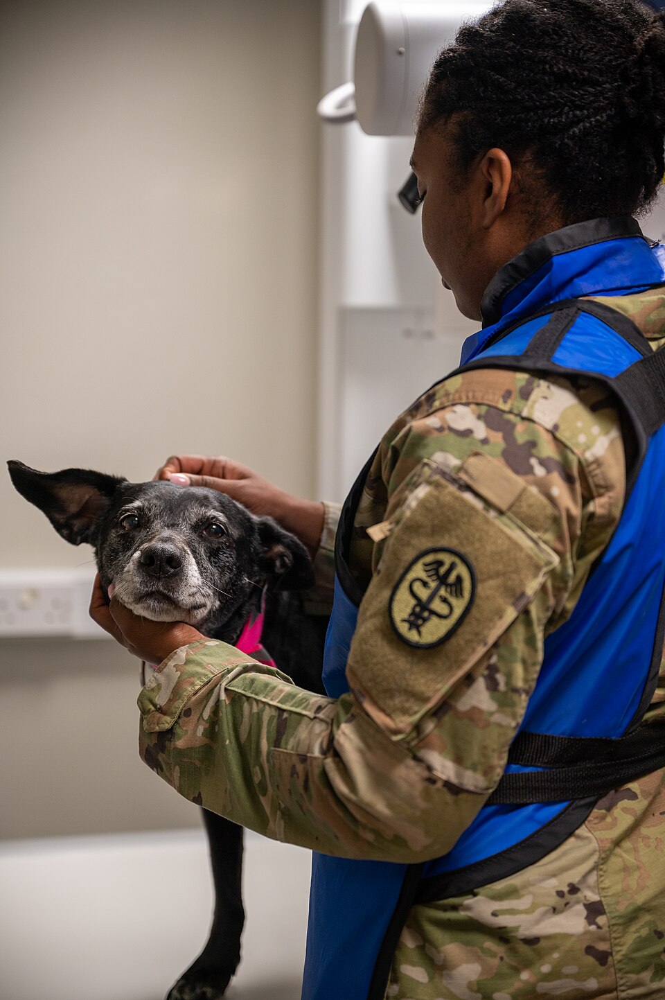
Time, distance and shielding
Dose depends on how long a person is exposed, how much radiation the source produces, how far away the person is and what shielding stands between them. The main controls in a veterinary clinic are to shorten exposure, increase distance and use proper shielding.2
| Method | What it means | Example in practice |
|---|---|---|
| Time | Less exposure time, fewer exposures | Position carefully the first time so you do not need retakes; use a calibrated technique chart |
| Distance | Stand as far from the source and patient as you can | Leave the room during the exposure; if you must stay, step back from the table |
| Shielding | Lead between you and the radiation | Apron with 0.5 mm lead, thyroid shield, full-coverage gloves, lead glasses for large animal work |
| Collimation | Limit the beam to the area of interest | Collimate to about 1 cm outside the patient's edges; never rely on digital cropping |
| Hands-free restraint | Fewer people in the room | Sandbags, tape, foam wedges, troughs, sedation |
Sources: Merck Veterinary Manual; Today's Veterinary Nurse.123
The inverse square law
The exposure rate from a source falls with the square of the distance. Double the distance and the exposure rate drops to one fourth.4 Small moves matter: stepping from 1 m to 2 m from the source cuts the exposure rate to one fourth.
Personal protective equipment
Radiology PPE requirements vary by state. Best practice is a lead gown containing 0.5 mm of lead that wraps halfway around the wearer and covers from the thyroid to the knees, a lead thyroid shield and full-coverage (360 degree) lead gloves.3 Lead-impregnated glasses are recommended too, especially for large animal radiography, where exposures can be high and the beam is often horizontal.1
Know the limit of PPE. Lead aprons and gloves reduce scatter by a factor of about 1,000 but the primary beam by only about 10. Leaded gloves must never be placed in the primary beam.1 If you can see your glove on the image, your hand was in the primary beam.
Dosimetry badges
Anyone who takes radiographs should be monitored. Badge reports help find and correct conditions that cause excess exposure, and they give a record if a question later arises about whether a medical condition is linked to radiation at work.1 Common devices are the film badge, processed by a vendor that keeps a permanent record, and the thermoluminescent dosimeter (TLD), a lithium fluoride chip that gives off light in proportion to its dose when heated. TLDs are commonly used as extremity dosimeters, for example on the hands.4
If one badge is assigned, wear it on the collar, outside the lead apron. If two are assigned, the second goes at the waist under the apron.4 Badge reports subtract background radiation.4 A badge only measures; it does not protect.
Units and dose limits
The rem and its SI unit, the sievert (Sv), describe biological risk. One sievert equals 100 rem, so 1 rem equals 10 millisieverts (mSv).4 Federal and state authorities set maximum limits for whole-body and extremity exposure.1 The federal numbers below are from the Nuclear Regulatory Commission rule in 10 CFR Part 20. A training handout from LSU Health states that state and NRC rules both allow 5,000 mrem (5 rem) a year.4
| Who or what | Limit |
|---|---|
| Adult, whole body (total effective dose equivalent) | 5 rem (0.05 Sv, 50 mSv) per year |
| Lens of the eye | 15 rem (0.15 Sv) per year |
| Skin or any extremity (hands, feet) | 50 rem (0.5 Sv) per year |
| Embryo or fetus of a declared pregnant worker | 0.5 rem (5 mSv) for the entire pregnancy |
| Minors (under 18) | 10 percent of the adult limits |
Sources: 10 CFR 20.1201, 20.1207 and 20.1208.567
Limits are a ceiling, not a target. ALARA applies at every dose, however far below the limit.1
Procedure: a safe exposure, step by step
- Confirm the request and the views needed, so nothing is repeated.
- Ask whether the patient can be sedated. Sedation or short-acting anesthesia is often needed, reduces manual restraint and leads to fewer unacceptable radiographs.1
- Position with sandbags, tape, foam pads and troughs so fewer people need to stay in the room.13
- Measure the patient and set the technique from a calibrated technique chart.3
- Collimate to about 1 cm outside the patient's edges. If the whole detector is exposed, the beam is probably undercollimated.1
- Anyone who must stay puts on apron, thyroid shield and gloves, with the badge at the collar outside the apron, and stands as far from the beam as possible.34
- Check that no hand, gloved or not, is in the collimated field. Then expose.
- Review the image before moving the patient, so a needed retake is done once and correctly.
Flowchart: who stays in the room?
Some states do not allow manual restraint except in defined circumstances.1 Know your state's rule before you hold.
Species differences
- Dogs and cats: most exams can be done with sedation and positioning aids in about the same time as manual restraint, and the patient is less likely to injure staff or itself.1
- Horses: upper limb, cervical spine and skull studies are likely to expose the upper body and head of anyone holding the detector or the horse. The beam is often horizontal and exposures can be high, so lead glasses are recommended.1
- Cats treated with radioactive iodine (I-131) for hyperthyroidism: the cat itself becomes a source. It is isolated, and its urine, feces and bedding are handled and disposed of with care.2
Calculation
Formula: new exposure = old exposure x (old distance / new distance) squared.4
Example 1. A technician receives 16 mR per hour of scatter at 1 m from the table. She steps back to 2 m. New exposure = 16 x (1/2) squared = 16 x 1/4 = 4 mR per hour.
Example 2. She instead moves from 1 m to 3 m. New exposure = 16 x (1/3) squared = 16 / 9 = about 1.8 mR per hour.
Example 3, moving closer. She moves from 2 m to 1 m. New exposure = 4 x (2/1) squared = 4 x 4 = 16 mR per hour. Halving the distance quadruples the exposure.
Unit check. A badge report of 200 mrem is 0.2 rem, or 2 mSv (1 rem = 10 mSv).4
Safety, legal and client education
- Registration and inspection. The FDA regulates the design and radiation control of x-ray equipment. Individual states regulate how x-ray machines are used, including personnel qualifications and quality control.8 Registration, inspection and record rules therefore come from your state radiation control program and differ by state. Keep machine registration and dosimetry records where an inspector can find them.
- Pregnant staff. Personnel who are pregnant or under 18 should not be directly involved in taking radiographs whenever possible.1 Once a worker declares her pregnancy, the fetal limit is 0.5 rem (5 mSv) for the whole pregnancy, and the employer should avoid large month-to-month swings in her dose.6
- Clients. When an owner asks to stay and hold, explain that sedation and positioning aids give a better image with fewer retakes and keep people out of the radiation field.
Common mistakes and exam traps
- Trap: "Lead gloves make it safe to hold the leg in the beam." They cut the primary beam only about tenfold. Hands stay out of the beam.1
- Trap: doubling the distance halves the dose. It cuts it to one fourth.4
- Trap: wearing the only badge under the apron. That reads the shielded dose and misses the thyroid and eyes. Wear it at the collar, outside.4
- Trap: cropping a digital image instead of collimating. Cropping changes the picture, not the dose.1
- Trap: confusing the limits. Whole body 5 rem, lens 15 rem, extremities 50 rem, fetus 0.5 rem per pregnancy.56
- Trap: thinking the primary beam is the main hazard to staff. The usual staff dose is scatter from the patient.1
Memory aids
TDS: Time, Distance, Shielding, the three main controls for staff dose (memory aid).
Twice as far, a quarter of the dose: doubling your distance cuts exposure to one fourth (memory aid).
5, 15, 50, 0.5: annual limits in rem for whole body, lens of the eye and extremities, then the fetal limit for a whole declared pregnancy (memory aid).
Badge at the neck: one badge, collar level, outside the apron, closest to the unshielded thyroid and eyes (memory aid).
1,000 and 10: lead cuts scatter about a thousandfold but the primary beam only about tenfold (memory aid).
Quick review
- ALARA: the lowest dose that still gives a diagnostic image. No dose is completely safe.
- Control staff dose with time, distance and shielding, plus tight collimation and hands-free restraint.
- Scatter from the patient is the main source of staff exposure.
- Inverse square law: 2 times the distance gives 1/4 the exposure; 3 times gives 1/9.
- PPE: 0.5 mm lead apron, thyroid shield, full-coverage gloves, lead glasses for large animal work. Never in the primary beam.
- One badge at the collar outside the apron; a second at the waist under it.
- Limits: 5 rem whole body, 15 rem lens, 50 rem extremities per year; 0.5 rem fetus per pregnancy; minors 10 percent.
- Pregnant and under-18 staff should avoid taking radiographs whenever possible.
- States regulate x-ray machine use and inspect; the FDA regulates the equipment itself.