Introduction
Fecal examination is one of the most common tests a veterinary technician runs. It is cheap, it does not hurt the patient, and it finds most of the intestinal worms and protozoa seen in dogs, cats and livestock.1 The technician usually does the whole job: telling the client how to collect the sample, choosing and setting up the test, scanning the slide and recognizing the egg or cyst. The veterinarian then makes the diagnosis and picks the treatment.
Many of these parasites can also infect people. Good technique protects the patient, the family and the clinic staff. This guide covers the main fecal tests, flotation solutions and their specific gravity, how to identify the common eggs and cysts, and the zoonotic points you will explain to clients.
Key facts
- Use the freshest sample you can get. If it cannot reach the lab within 2 hours, refrigerate it. A sample left at room temperature for more than 6 hours is unsuitable.1
- Centrifugal flotation is more sensitive than simple (passive) flotation. CAPC recommends it for routine screening.12
- CAPC advises a flotation solution with a specific gravity (SG) of 1.18 to 1.27: sodium nitrate is 1.18 to 1.20 and Sheather's sugar solution is 1.27.2
- Zinc sulfate at SG 1.18 with centrifugation is the best flotation for Giardia cysts. Stronger solutions can distort the cysts.116
- The direct smear finds motile trophozoites, but only in very fresh feces (within 20 minutes of passing).1
- Sedimentation is the method of choice for operculate eggs such as fluke eggs. The Baermann test is the method of choice for nematode larvae such as lungworms.1
- One negative fecal does not rule out parasites. Merck advises three samples, on consecutive days or over 7 to 10 days.1
- Roundworms (Toxocara) and hookworms (Ancylostoma) are zoonotic. Toxocara causes larva migrans syndromes in children; animal hookworms are the most common cause of cutaneous larva migrans.34
How the VTNE tests this
- Choose the test: which fecal method suits a given patient or suspected parasite. Domain 4 Task C lists parasitology among the laboratory tests a technician performs, and K25 covers laboratory principles, procedures and methods, including parasitology.15
- Choose the solution: name the flotation solution and its specific gravity for a task, such as zinc sulfate for Giardia.
- Identify: name the parasite from a description of an egg, cyst, oocyst or tapeworm segment.
- Troubleshoot: explain a false negative, such as an old sample, too little feces or a slide read too late.
- Educate: give zoonosis and hygiene advice. K36 covers public health, including infection control and zoonosis.15
Core concepts
Why flotation works
Flotation separates parasite stages from fecal debris by density. A flotation solution is salt or sugar dissolved in water. The more you dissolve, the higher the density, measured as specific gravity (SG). Water has an SG of 1.000. Eggs and cysts that are less dense than the solution rise to the top. Heavier debris sinks.2 A centrifuge applies far more force than gravity, so separation is faster and more complete.2
Choosing the SG is a trade off. A denser solution floats heavier eggs, but it also floats more debris and can collapse thin-walled stages, such as Giardia cysts, so they are hard to recognize.2 Whipworm eggs are fairly dense (average SG 1.15), so they need proper technique with centrifugation to be recovered.5
Viscosity matters too. Sheather's sugar solution is more viscous, works well in the centrifuge and holds the coverslip on the tube better. Salt solutions dry fast and form crystals on the slide, so read salt preparations quickly.2 Sugar preparations can be kept in the refrigerator for a few days if you need to recheck a slide.1
Shedding is patchy
Parasites do not shed eggs or cysts at a steady rate. A single negative result does not rule out parasites in a sick animal.1 Some parasites also cause signs before eggs appear. The whipworm Trichuris vulpis has a prepatent period (time from infection to first eggs in feces) of 74 to 90 days and may shed eggs on and off.5
False positives
A dog that eats feces (coprophagia) or a pet that hunts can pass eggs, cysts or larvae from another animal for several days without being infected.1 Raccoon roundworm eggs (Baylisascaris) found in a dog's feces after it eats raccoon feces should disappear within 1 to 2 days, so a repeat sample helps confirm a true infection.9
Procedure or technique
Sample collection and client instructions
- Give the client a leakproof, sealable container with the patient's name on it.1
- Dog owners should collect the stool right after the dog passes it, so it is certain to come from their dog. Cat owners should clean the litter box and collect the next stool.1
- Bring it in within 2 hours or refrigerate it. Do not use stool that has sat on soil or grass for a long time, because free-living nematodes can invade it.1
- At the bench, look at the stool first. Note blood, mucus, whole worms or tapeworm segments.2
Centrifugal flotation (the routine test)
- Weigh or measure enough feces. CAPC advises at least 1 g of formed feces, 2 g if soft, 4 g if slurry-like and 6 g or more if liquid. A fecal loop sample is too small and can give a false negative.2 Merck uses about 1 teaspoon (3 to 5 g).1
- Mix the feces with flotation solution and strain it through cheesecloth or a tea strainer into a centrifuge tube.12
- Balance the tubes in opposite buckets. With a swinging-bucket centrifuge, top up to a domed meniscus, place the coverslip and spin. With a fixed-angle centrifuge, spin the tube nearly full, then top it up and add the coverslip after it stops, and let it stand a few minutes.2
- Spin for about 10 minutes. CAPC describes bringing a swinging-bucket rotor up slowly to a maximum of 800 rpm; Merck gives 650 g for 10 minutes. Follow your centrifuge maker's settings.12
- Let the rotor stop on its own. Lift the coverslip, place it on a slide and scan the entire coverslip in a pattern with the 10x objective.12 Focusing on a small air bubble helps you find the right focal plane.2
Simple (passive) flotation
Commercial kits use a chamber and a filter insert. Mix a small amount of feces with solution, fill to a positive meniscus, add a coverslip and wait 5 to 10 minutes before reading.1 It is quick but less sensitive. In one CAPC class exercise on a hookworm-positive dog, about 25% of students found eggs by direct smear, about 70% by passive flotation and 100% by centrifugal flotation.2
Direct smear
Mix a speck of feces on an applicator stick into a drop of saline on a slide and add a coverslip. The film should be thin enough to read print through it. Turn the condenser down to boost contrast.1 Use it for motile trophozoites such as Giardia and Tritrichomonas blagburni, on feces no more than 20 minutes old. Because so little feces is examined, false negatives are common.1
Simple sedimentation
Mix 1 to 2 g of feces with water, strain it into a tube and let it stand 5 minutes. Pour off the fluid, refill with water, resuspend and let it stand again. Repeat a third time, then examine drops of the sediment.1 Use saline instead of water if the blood fluke Heterobilharzia americana is suspected, because water makes its eggs hatch.1
Baermann technique
Set a glass funnel with tubing and a clamp in a rack. Half fill it with warm tap water (comfortable to touch). Wrap 10 to 20 g of fresh feces in a double layer of cheesecloth, hang it in the water on an applicator stick and leave it for at least 8 hours. Collect the first fluid into a 15 mL tube, spin it, pour off the top and examine the sediment.1 Live larvae swim out of the feces and sink. A drop of dilute iodine kills them so they can be identified.1
| Technique | Best for | Solution and key detail |
|---|---|---|
| Centrifugal flotation | Routine screening; most helminth eggs, protozoal cysts and oocysts | SG 1.18 to 1.27 (sodium nitrate 1.18 to 1.20, Sheather's sugar 1.27); spin about 10 min |
| Zinc sulfate centrifugal flotation | Giardia cysts | Zinc sulfate, SG 1.18; less distortion of cysts |
| Simple (passive) flotation | Quick in-clinic kit screen | Same solutions; stand 5 to 10 min; lower sensitivity |
| Direct saline smear | Motile trophozoites (Giardia, Tritrichomonas) | Saline; feces under 20 min old; small sample, many false negatives |
| Simple sedimentation | Operculate eggs (flukes, Diphyllobothrium, Spirometra), Physaloptera, acanthocephalans | Water (saline for Heterobilharzia); settle and decant 3 times |
| Baermann | Nematode larvae: Aelurostrongylus, Angiostrongylus, Crenosoma, Strongyloides | Warm water; 10 to 20 g fresh feces; at least 8 h |
Source: Merck Veterinary Manual, Parasitology in Veterinary Practice; CAPC, Microscopic Fecal Exam Procedures.12
Recognizing the common eggs and cysts
Scan at 10x, then move up to confirm detail. Compare size against a calibrated eyepiece if your microscope has one. The descriptions below are the features exam questions use.
| Parasite | What you see | Host | Zoonotic? |
|---|---|---|---|
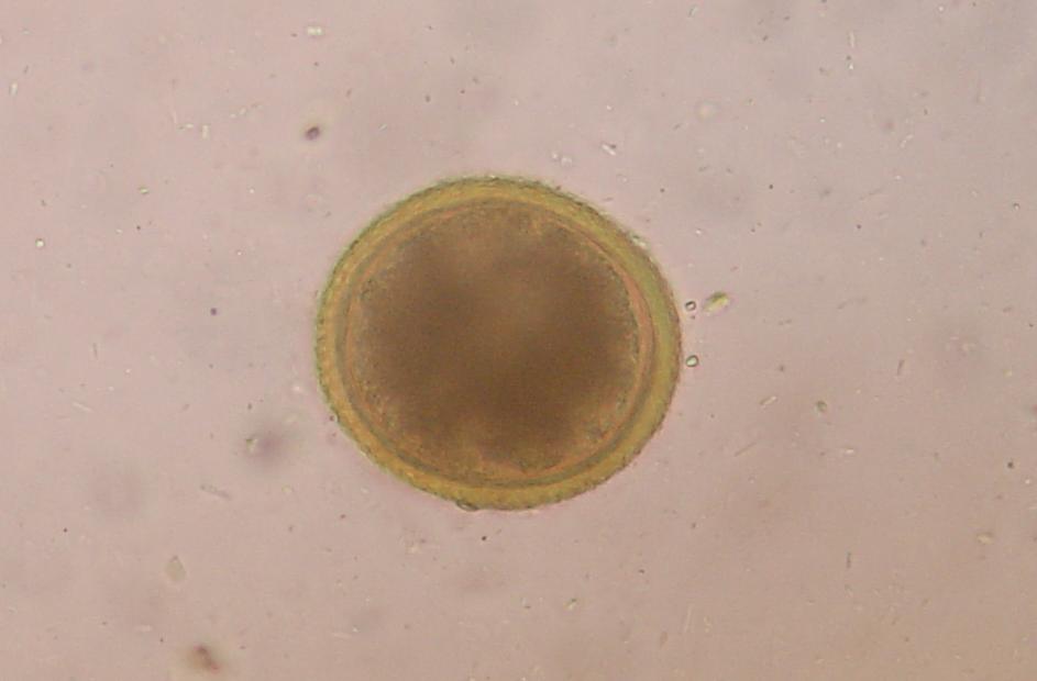
| Toxocara canis, T. cati (roundworms) | Round egg with a rough, pitted shell and a dark single cell; T. canis about 80 to 90 by 75 µm | Dog (T. canis), cat (T. cati) | Yes: visceral, ocular and neural larva migrans |
| Toxascaris leonina (roundworm) | Oval egg, smooth outer shell, wavy inner surface, lighter embryo; 75 to 85 by 60 to 75 µm | Dog and cat | Not the species of public health concern |
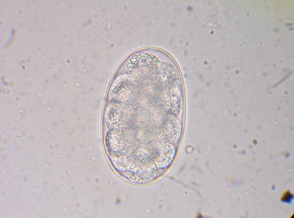
| Ancylostoma (hookworm) | Oval, thin-walled egg with a morula (cluster of 2 to 8 cells); 52 to 79 by 28 to 58 µm | Dog and cat | Yes: cutaneous larva migrans |
| Trichuris vulpis (whipworm) | Barrel or football-shaped, yellow-brown egg with a plug at each end (bipolar plugs); about 79 by 38 µm | Dog (rare in US cats) | Not considered zoonotic at this time (CAPC) |
| Taenia (and Echinococcus) | Single taeniid egg holding a hexacanth (six-hooked) embryo; the two genera look the same | Dog and cat | Echinococcus: yes, handle with great care |
| Dipylidium caninum (flea tapeworm) | Cucumber-seed segment (proglottid) 10 to 12 mm, two genital pores; egg packets of 25 to 30 eggs | Dog and cat | Occasionally, mainly infants who swallow an infected flea |
| Giardia | Cyst: clear, oval, 8 to 12 by 7 to 10 µm, 2 to 4 nuclei. Trophozoite: pear-shaped, 2 nuclei, motile | Dog and cat | Possible; spread from pets to people appears rare |
| Coccidia (Cystoisospora) | Oocysts, unsporulated (not yet infective) when passed | Dog and cat, each host-specific | Canine Cystoisospora: not known to be zoonotic |
| Toxoplasma gondii | Small unsporulated oocyst, about 10 by 12 µm, in cat feces only | Cat (definitive host) | Yes: pregnant and immunocompromised people |
Sources: CAPC guidelines for ascarids, hookworms, Trichuris vulpis, Dipylidium caninum, Echinococcus, Giardia, coccidia and Toxoplasma gondii; Merck Veterinary Manual roundworm and tapeworm chapters.3456789121314
Notes on tapeworms
Fecal flotation is poor at finding tapeworms. Dipylidium segments must break open to release egg packets, and they are not spread evenly through the stool. In one shelter study of more than 40 cats, flotation found no eggs in any cat that had adult Dipylidium.6 Owners often see the segments instead, moving near the anus, on bedding or on the stool. Crush a segment in water or saline and look for egg packets under the microscope.6 Taenia and Echinococcus eggs look identical; PCR is needed to tell them apart.12
Flowchart: which fecal test?
Species differences
- Dogs: transplacental transmission (from the mother before birth) is an important route of Toxocara canis infection, so puppies can shed eggs young.3 Whipworm eggs with bipolar plugs are a dog finding.
- Cats: whipworm infection is rare in US cats. An egg with bipolar plugs in cat feces is more likely a capillarid such as Eucoleus aerophilus.5 Only cats and other felids shed Toxoplasma oocysts.14 The Baermann test is the method of choice for the cat lungworm Aelurostrongylus abstrusus.1
- Coccidia: Cystoisospora species are strictly host-specific; canine coccidia do not infect cats.8
- Horses and ruminants: collect feces from the rectum with a glove when you can. For a herd, sample at least 10 animals. Quantitative fecal egg counts (FECs) measure eggs per gram (EPG). They are used to detect dewormer resistance and to guide selective deworming in horses.1 The McMaster slide is common in horses and small ruminants. Saturated salt (SG 1.20) is a cheap solution for production animals, and magnesium sulfate (SG 1.20) is preferred for pig feces.1
Calculation
McMaster fecal egg count. Mix 4 g of feces with 56 mL of sugar flotation solution (60 mL total). Each McMaster chamber holds 0.15 mL under the grid, so the two chambers hold 0.3 mL.1
Fraction examined: 60 mL ÷ 0.3 mL = 200, so the whole sample is 200 times what you counted. Divide by the 4 g of feces: 200 ÷ 4 g = 50. Each egg you count equals 50 eggs per gram.1
Example: a horse sample shows 9 strongyle eggs in chamber 1 and 7 in chamber 2.
(9 + 7) eggs × 50 EPG per egg = 800 EPG.
Merck notes there is no agreed cut-off, but thresholds of 250 EPG or more are often used in horse strongyle control programs, so report this result to the veterinarian.1
Safety, legal and client education
- Roundworms: Toxocara eggs need 2 to 4 weeks in the environment before they are infective, but they then survive for years and common disinfectants do not kill them.3 Tell families to pick up feces right away on walks and daily from the yard, and to protect sandboxes, gardens and playgrounds from fecal contamination. Early, regular deworming of puppies and kittens cuts environmental contamination.3
- Hookworms: people are infected when larvae penetrate the skin, which causes itchy, winding skin tracks. Prompt removal of pet feces protects public areas.4 Hookworm eggs hatch in 24 to 72 hours on warm, moist soil.10
- Giardia: cysts are infective as soon as they are passed.7 Wear gloves and clean up promptly.
- Toxoplasma: remove cat feces from the litter box every day. Oocysts need 1 to 5 days to become infective. Pregnant and immunocompromised people need extra care.14
- Dipylidium: the pet is infected by swallowing an infected flea while grooming, so flea control is part of prevention.6 People, particularly infants, are occasionally infected the same way.12
- Echinococcus: taeniid eggs could be Echinococcus, which is zoonotic with a low infective dose. CAPC advises extreme caution when processing samples from suspect dogs, such as hunting dogs with access to offal.13
- In the lab: wear gloves, keep food and drink out of the lab, balance the centrifuge, and dispose of fecal samples as biohazard waste under your clinic's protocol.
Common mistakes and exam traps
- Using a fecal loop sample: too little feces gives false negatives. Use at least 1 g of formed stool.2
- Reading salt preparations late: salt solutions dry and crystallize on the slide. Read them promptly.2
- Looking for trophozoites in old or refrigerated feces: they die and become unrecognizable fast. Use feces under 20 minutes old.1
- Expecting flotation to find everything: trophozoites, nematode larvae, spirurid eggs, acanthocephalan eggs and operculate eggs are not reliably found by flotation.1
- Using a Giardia ELISA to check treatment: it can stay positive long after treatment, so it is not suitable for post-treatment monitoring.17
- Mixing up the two roundworms: pitted shell means Toxocara; smooth shell means Toxascaris.9
Quick review
- Fresh feces: within 2 hours or refrigerate; over 6 hours at room temperature is unsuitable.
- Centrifugal flotation is the routine screen and beats simple flotation.
- Flotation SG 1.18 to 1.27: sodium nitrate 1.18 to 1.20, Sheather's sugar 1.27.
- Giardia cysts: zinc sulfate SG 1.18 with centrifugation.
- Trophozoites: direct saline smear on feces under 20 minutes old.
- Operculate (fluke) eggs: sedimentation. Nematode larvae: Baermann.
- Toxocara: round, pitted shell. Toxascaris: oval, smooth shell.
- Hookworm: oval, thin shell, morula of 2 to 8 cells.
- Whipworm: barrel-shaped with bipolar plugs; dense eggs, long prepatent period.
- Dipylidium: cucumber-seed segments and egg packets; spread by fleas.
- In a sick animal, check three samples (consecutive days or over 7 to 10 days) before ruling out parasites.
- McMaster (4 g in 56 mL): eggs counted in both chambers × 50 = EPG.