Introduction
Ruminant nursing is mostly herd work. On a dairy or beef call the technician restrains, draws blood, runs cowside tests, gives injections, drenches cows, feeds colostrum to calves and keeps the treatment record that decides when milk and meat can be sold. The disorders you meet most often cluster around calving: milk fever, ketosis and mastitis in the fresh cow, and failure of passive transfer in the newborn calf. Bloat can kill a cow within hours. This guide covers the rumen basics behind these problems, what each condition looks like, the technician's tasks, Beef Quality Assurance (BQA) injection rules and how withdrawal times keep drug residues out of food.
Key facts
- Milk fever (parturient paresis) has three stages: standing and excitable, down but sternal, then lateral and comatose.2
- The standard treatment for a recumbent cow is 500 mL of 23% calcium gluconate IV, given slowly over 10 to 20 minutes while the heart is monitored.2
- Ketosis (hyperketonemia) threshold: blood beta-hydroxybutyrate (BHB) of 1.2 mmol/L. Most cases occur in the first 2 weeks of lactation.3
- The California Mastitis Test (CMT) mixes equal amounts of foremilk and reagent in a four-cup paddle. Gel formation means more leukocytes (somatic cells).5
- Calves need at least 3 L of high-quality colostrum (IgG above 50 mg/mL) within 6 hours of birth, or 4 L within 6 hours.6
- BQA: give injections in the neck, in front of the shoulder, no more than 10 mL per site, with sites at least 5 inches apart.9
- Failure to observe the correct withdrawal time is the most common cause of violative drug residues in food.11
How the VTNE tests this
The VTNE outline lists disease control and prevention techniques, including vaccination and herd health, as a knowledge area, and asks technicians to give medications by the appropriate routes.16 Ruminant items tend to test one practical point at a time:
- Recognize the stage of milk fever from a short description, and name the monitoring task during IV calcium (Domain 5 Task B; K38).
- Read a CMT result, or put CMT and milk culture steps in the right order.
- Choose the BQA injection site and maximum volume per site (Domain 1 Task D).
- Name the resource for extralabel withdrawal times (FARAD) and explain why label withdrawal times matter.
- Calculate colostrum volume, doses or the number of injection sites a dose needs.
Core concepts
The rumen
Cattle, sheep and goats have a three-compartment forestomach (rumen, reticulum and omasum) and a true stomach, the abomasum. Microbes in the rumen ferment feed and produce volatile fatty acids (VFAs): acetic, propionic and butyric acid.14 Fermentation also makes a lot of gas, which the cow must belch up (eructate). Anything that stops eructation, or traps gas in foam, leads to bloat. A sudden diet of grain does the opposite kind of damage: lactic acid builds up and rumen pH falls to 5 or below, which kills the normal fiber-digesting microbes and stops rumen motility.15
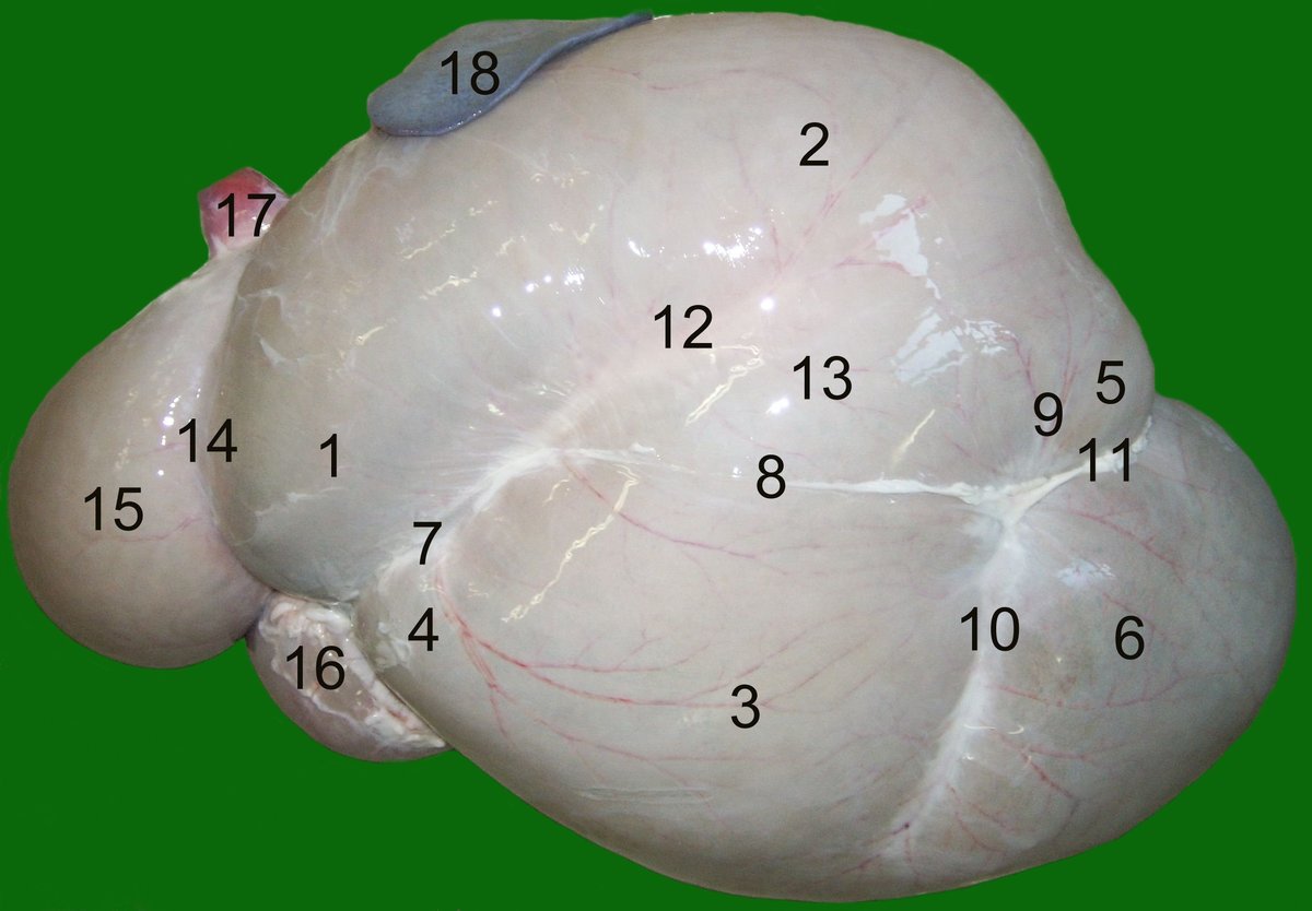
Bloat
Bloat is overdistention of the rumen and reticulum with fermentation gas. In frothy (primary) bloat the gas is trapped in a stable foam. In free-gas (secondary) bloat the gas is separate from the ingesta, usually because something blocks eructation, such as an esophageal foreign body.1 The left flank swells first, and in pasture bloat the left paralumbar fossa can bulge above the line of the spine. Death can occur within 1 hour of starting to graze but is more common 3 to 4 hours after signs begin. Passing a stomach tube releases gas and relieves free-gas bloat; it does not relieve frothy bloat, which needs an antifoaming agent.1
Milk fever (parturient paresis)
At the start of lactation a dairy cow's daily calcium loss jumps from about 10 g to about 30 g. Normal blood calcium is about 8.5 mg/dL (2.12 mmol/L) at the low end; in milk fever it usually drops below 6.0 mg/dL (1.5 mmol/L). Jersey and Guernsey cows are more often affected. Signs usually appear within 48 hours of calving.2
- Stage 1: the cow is standing but excitable and hypersensitive, with fine tremors, ear twitching and shuffling of the hind feet.
- Stage 2: she cannot stand but stays sternal. Look for a dull attitude, dry muzzle, low temperature, cold extremities, a fast heart with quiet heart sounds, bloat, and the head tucked into the flank. An S-shaped curve may be seen in the neck.
- Stage 3: she lies flat on her side, loses consciousness and can bloat badly. The heart rate can approach 120 beats per minute. Untreated, she may survive only a few hours.2
Ketosis
Hyperketonemia is a raised level of ketone bodies (acetone, acetoacetate and BHB) in the blood. It is a disease of early lactation: all cows are at risk for the first 6 weeks, and most cases occur in the first 2 weeks. Signs are vague: poor appetite, falling milk yield, loss of body condition and firm, dry feces. A few cows show nervous ketosis, with abnormal licking, incoordination, bellowing or aggression.3 Herds screen fresh cows with handheld blood BHB meters, milk tests or urine dipsticks.3
Mastitis and somatic cells
Mastitis is inflammation of the mammary gland, usually from infection. When bacteria invade a quarter, leukocytes (white blood cells) pour into the milk. These leukocytes make up most of the somatic cell count (SCC). An SCC of 200,000 cells/mL or more in a cow indicates a high likelihood of infection.4 Clinical mastitis, with watery, thick or ropy milk, is only about 20% of the mastitis in a dairy herd. Most infected quarters give normal-looking milk, which is why a cowside screen such as the CMT is useful.5
| Condition | Signs you may see | Technician tasks |
|---|---|---|
| Frothy bloat | Sudden left flank swelling on lush legume pasture; foamy rumen contents | Remove from pasture, call the veterinarian, prepare antifoaming agent and stomach tube |
| Free-gas bloat | Left flank swelling; gas comes off when a tube is passed | Prepare stomach tube; check for choke (esophageal obstruction) |
| Milk fever | Within 48 h of calving: tremors, then sternal with head in flank, cold ears, then lateral | Draw a pretreatment blood sample; set up IV calcium; monitor heart during infusion; keep cow sternal |
| Ketosis | Early lactation; off feed, less milk, weight loss, dry feces; sometimes licking or aggression | Run blood BHB, milk or urine cowside test; drench propylene glycol as ordered |
| Subclinical mastitis | Normal-looking milk; high SCC | Run CMT on each quarter; collect aseptic sample for culture as ordered; record quarter results |
| Failure of passive transfer | Calf that nursed poorly or late; low serum total protein | Feed or tube colostrum on time; draw blood for serum IgG or total protein |
Source: Merck Veterinary Manual pages on bloat, parturient paresis, hyperketonemia, mastitis and calf feeding; MU Extension CMT guide.12356
Procedure or technique
Assisting with IV calcium for a down fresh cow
- Keep the cow sternal. Recumbent cows are at very high risk of muscle and nerve damage, so treatment must be prompt.2
- Draw a pretreatment blood sample before any calcium is given. If the cow does not respond, this sample can still be sent for calcium testing. A sample taken after treatment is useless for diagnosis.2
- Prepare the jugular site aseptically. The jugular is the preferred vein. The mammary vein is prone to thrombosis and phlebitis and is used only when neither jugular is available.2
- Give the calcium slowly, over 10 to 20 minutes, while listening to the heart or feeling the pulse. IV calcium briefly raises blood calcium to nearly twice the upper normal limit, which can cause a fatal arrhythmia. If the rhythm becomes irregular or the heart slows sharply, stop the infusion and tell the veterinarian.2
- Watch for a good response: the heart rate falls, the pulse gets stronger, muscle tremors appear and the cow tries to rise. About 75% of recumbent cows rise within 2 hours.2
A cow that is still standing (stage 1) is usually given oral or subcutaneous calcium instead.2
Running a California Mastitis Test
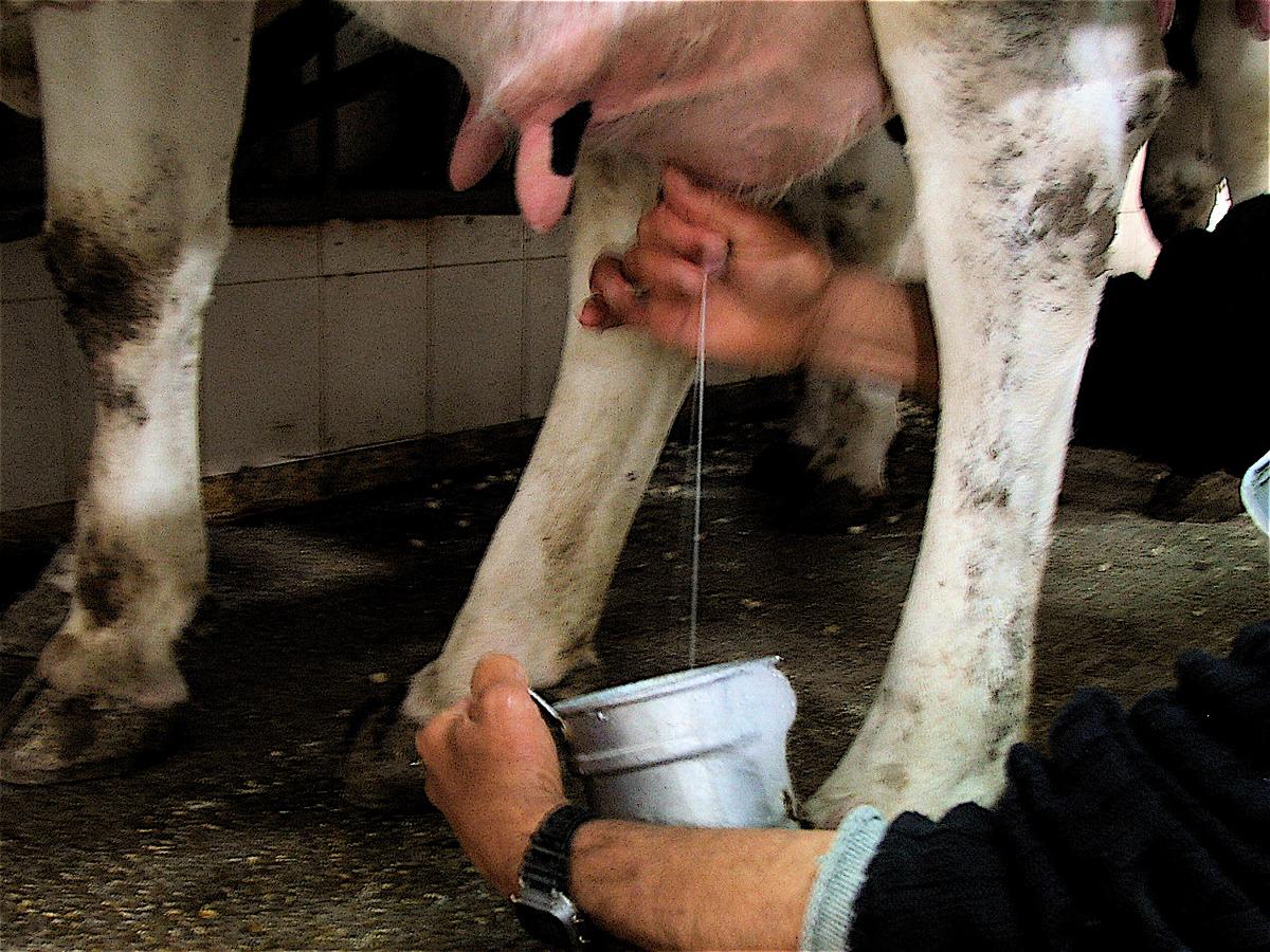
- Strip the first stream or two of foremilk from each quarter into its own cup of the four-cup paddle. Milk drawn later has fewer leukocytes.5
- Tilt the paddle so each cup holds about 1/2 teaspoon. Add an equal volume (about 1/2 teaspoon) of licensed CMT reagent.
- Rotate the paddle to mix. Read color and gel formation within 10 to 15 seconds.
- Score each quarter and record it by quarter (left front, right front, left rear, right rear).5
The CMT shows inflammation but does not identify the bacteria; that needs a laboratory culture.5
Colostrum management for calves
Timely feeding of enough high-quality colostrum is the single most important practice for newborn calf health. Feed at least 3 L of high-quality colostrum (IgG above 50 mg/mL) within 6 hours of birth, then a second feeding at 8 to 12 hours. An alternative plan is 4 L (about 1 gallon) within 6 hours. The first feeding matters most because the gut closes to antibody absorption quickly.6 Passive transfer is checked later on a blood sample (serum IgG or serum total protein).
| Category | Serum IgG (mg/mL) | Serum total protein (g/dL) | Target share of calves |
|---|---|---|---|
| Excellent | 25.0 or more | 6.2 or more | More than 40% |
| Good | 18.0 to 24.9 | 5.8 to 6.1 | About 30% |
| Fair | 10.0 to 17.9 | 5.1 to 5.7 | About 20% |
| Poor | Below 10.0 | Below 5.1 | As few as possible |
Source: Merck Veterinary Manual table of recommended criteria for passive transfer in dairy calves, modified from Godden, Lombard and Woolums 2019.7 The Poor row is everything below the Fair cut-offs.
Disbudding, dehorning and castration
Disbudding destroys the horn-producing cells (corium) of the horn bud before the bud attaches to the skull. At about 2 months of age the bud attaches to the frontal bone over the frontal sinus, and later the sinus extends into the horn, so dehorning older cattle can open the sinus. Hot-iron and caustic paste methods destroy the corium; a complete ring of hair around the bud must be included to stop regrowth. Caustic paste can burn the surrounding skin or the eyes if it runs. The cornual nerve, a branch of the trigeminal nerve, supplies the horn, and a cornual nerve block with local anesthetic desensitizes the area.8 The technician restrains the head, clips and preps the site, draws up the local anesthetic and analgesic ordered, and watches for bleeding afterward.
When calves are castrated with rubber bands, the clostridial vaccine program should include Clostridium tetani (tetanus) protection.10
Vaccination and BQA injection technique
Modified live vaccines (MLV) give stronger, longer-lasting immunity and may not need a booster, but they must be mixed on the farm and used right away. Killed vaccines need a booster for optimal protection, are safe for pregnant animals and partly used vials can be stored.10 MLV starts to lose effect about an hour after mixing, so mix only what you will use within an hour, keep it cool and out of sunlight, and never clean MLV syringes with disinfectant (use hot water).9
BQA rules protect the expensive cuts of meat. Give all injections in front of the shoulder, using the triangle of neck muscle for both IM and SC products. Use the SC route when the label allows. Never inject into the top butt or top of the rump. Inject straight in for IM; tent the skin for SC. Give no more than 10 mL per site, keep sites at least 5 inches apart and avoid wet or manure-covered skin.9 Injection-site lesions in cow carcass rounds have fallen sharply since the 1998 and 2000 audits, and the authors credit BQA education and training.17
Flowchart: the down cow
A "downer cow" is one that cannot rise and stand for at least 12 to 24 hours. Whatever the first cause, a cow down for more than 24 hours develops pressure damage to hind-limb muscles and limb nerves, especially on a hard surface.13 Your first jobs are to keep her safe, sternal and comfortable while the veterinarian works out the cause.
Nursing care for a downer cow, per the Merck manual: provide a deep sand bed (more than 25 cm) or at least 10 inches of dry straw, keep her hydrated and give pain relief as ordered so she keeps eating. If she needs help rising, an assistant grabs the root of the tail with both hands; grabbing any other part of the tail can injure it. Hip clamps must not be too tight, and the cow is lifted slowly.13 Flotation tanks use lukewarm water, never hot or cold.13
Species differences
- Dog and cat: postpartum hypocalcemia in the bitch (eclampsia) usually strikes at peak lactation, 2 to 3 weeks after whelping, often in small-breed dogs nursing large litters. Panting and restlessness come first, then tremors and seizures. Calcium is again given IV slowly, over 10 to 30 minutes for 10% calcium gluconate, while the heart is monitored for bradycardia or arrhythmias.18 Dogs and cats have no rumen, so rumen bloat has no small animal equivalent.
- Cattle: milk fever, ketosis and mastitis are mostly dairy problems; Jersey and Guernsey cows are more often affected by milk fever.2 Bloat is mainly a cattle disorder.1
- Sheep and goats: bloat also occurs in sheep.1 Small ruminants share the same forestomach layout as cattle.14 Check every drug's label species: a cattle withdrawal time does not transfer to a goat.11
- Horse: horses are not ruminants.
Calculation
Worked example 1: how many injection sites? The veterinarian orders a product labelled at 1 mL per 100 lb SC for a 1,350 lb cow.
Dose = 1,350 lb x (1 mL / 100 lb) = 13.5 mL. The lb units cancel.
BQA limit is 10 mL per site. 13.5 mL / 10 mL per site = 1.35, so round up to 2 sites. Give about 6.75 mL in each, both in the neck, at least 5 inches apart.9
Worked example 2: calcium gluconate in one bottle. A 23% solution contains 23 g per 100 mL. 500 mL x (23 g / 100 mL) = 115 g of calcium gluconate. The mL units cancel. This standard 500 mL dose supplies 10.7 g of elemental calcium.2
Safety, legal and client education
- Withdrawal time is the time from the last dose until residues in meat or milk fall below the legal limit. The label withdrawal time applies only when the drug is given exactly as labelled. Changing the dose, route or frequency invalidates it.11
- Extralabel use in food animals is allowed only under the Animal Medicinal Drug Use Clarification Act (AMDUCA), and a small number of drugs are prohibited outright in food animals.11 For extralabel withdrawal recommendations, US veterinarians contact the Food Animal Residue Avoidance Databank (FARAD).12
- Mark treated cows clearly, record the drug, dose, route, site, date and withdrawal date, and tell the producer when milk can return to the tank.
- Read every label for dose, timing, route, withdrawal time, storage and expiry.9 Discard bent or burred needles.9
- Personal safety: work a down or excited cow from behind a barrier where possible, and never stand where a cow trying to rise can fall on you.
- Explain colostrum timing to producers: the first feeding in the first 6 hours does the most good.6
Common mistakes and exam traps
- Pushing IV calcium fast. The danger is a fatal arrhythmia; give it over 10 to 20 minutes and monitor the heart.2
- Drawing the diagnostic blood sample after the calcium. Collect it before treatment.2
- Expecting a stomach tube to fix frothy bloat. It relieves free-gas bloat; foam needs an antifoaming agent.1
- Thinking the CMT names the organism. It only shows inflammation; culture identifies the bacteria.5
- Injecting in the rump or top butt, or giving 20 mL in one site. BQA says neck, in front of the shoulder, 10 mL maximum.9
- Assuming the label withdrawal time still applies after an extralabel dose. It does not; the veterinarian sets a new one, often with FARAD's help.1112
Memory aids
Stand, Sternal, Side: the three stages of milk fever, from standing and excitable, to down but sternal, to lateral recumbency (memory aid).
3-6-2nd at 12: at least 3 L of good colostrum within 6 hours, with a second feeding by 8 to 12 hours (memory aid).
NECK 10-5: neck injections only, in front of the shoulder, 10 mL maximum per site, sites 5 inches apart (memory aid).
FARAD: Food Animal Residue Avoidance Databank, where veterinarians get extralabel withdrawal advice (acronym).
N, T, 1, 2, 3: CMT scores from negative to strong gel. Score 1 and above sits above 200,000 cells/mL (memory aid).
Quick review
- Ruminants have a rumen, reticulum and omasum plus a true stomach, the abomasum.
- Frothy bloat traps gas in foam; free-gas bloat is relieved by a stomach tube.
- Milk fever: within 48 hours of calving; stages are standing, sternal, lateral.
- Give 500 mL of 23% calcium gluconate IV over 10 to 20 minutes while monitoring the heart.
- Draw the blood sample before calcium is given.
- Ketosis threshold is blood BHB 1.2 mmol/L; treatment is an oral propylene glycol drench.
- SCC of 200,000 cells/mL or more suggests an infected quarter.
- CMT: foremilk plus equal reagent, swirl, read in 10 to 15 seconds; gel means leukocytes.
- Calves: 3 L of good colostrum within 6 hours (or 4 L), second feeding at 8 to 12 hours.
- Disbud before the horn bud attaches to the skull at about 2 months.
- BQA: neck only, 10 mL per site, 5 inches apart, SC when the label allows.
- Extralabel use voids the label withdrawal time; FARAD gives withdrawal advice.