Introduction
Some emergencies walk through the door again and again: the blocked male cat, the large dog with a swollen belly that retches but brings nothing up, the panting dog after a hot walk, the dog that will not stop seizing. Each one can kill within hours, and each one has a short list of jobs the technician can start at once while the veterinarian makes decisions.
This guide covers urethral obstruction and the hyperkalemia ECG, gastric dilatation and volvulus (GDV), oxygen supplementation, seizures and status epilepticus, diabetic ketoacidosis (DKA), heat stroke, dystocia, proptosis, and the monitoring sheet that ties ongoing care together.
Key facts
- In a blocked male cat, a heart rate below 120 beats/min together with hypothermia is 98% specific for potassium above 8.0 mmol/L and calls for immediate treatment.3
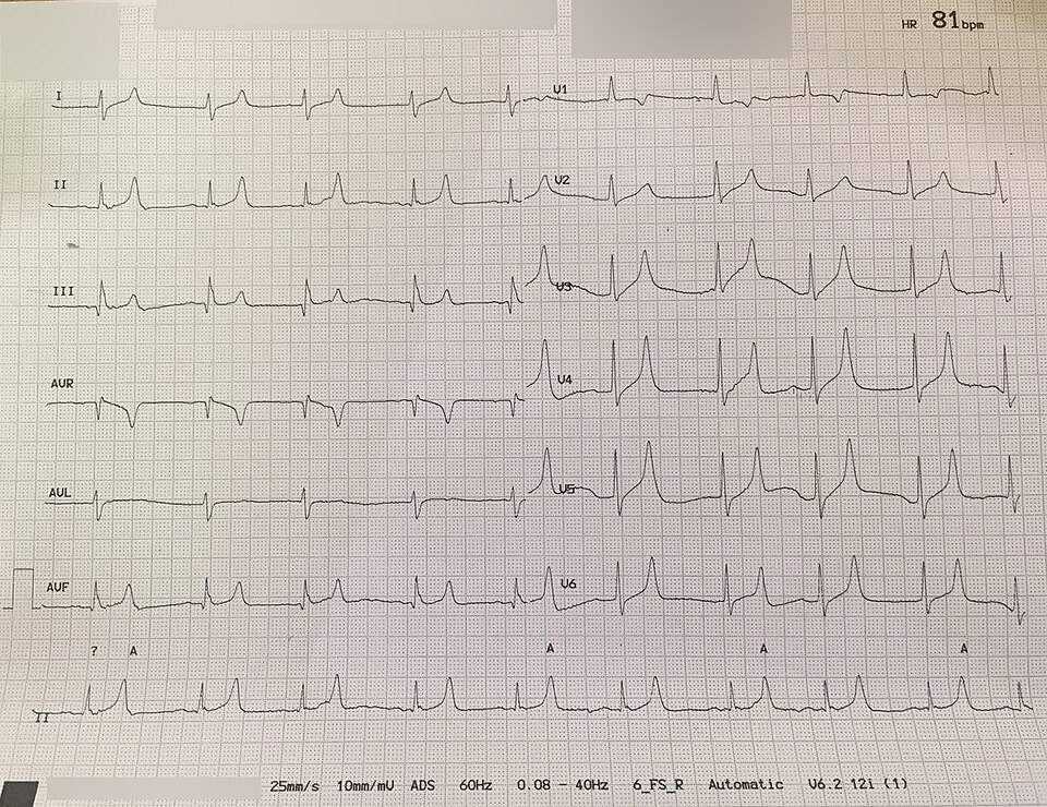
- Hyperkalemia ECG signs are small or absent P waves, a long PR interval, wide QRS complexes and tall, tented T waves, usually seen in lead II once potassium is above 7 mEq/L.2
- Calcium gluconate protects the heart within minutes but does not lower serum potassium.1
- A GDV patient needs one or more large-bore (16 to 18 gauge) catheters in a cranial vein, and stomach decompression runs at the same time as fluid resuscitation.4
- Status epilepticus is a seizure lasting more than 5 minutes, or repeated seizures without full recovery in between.37
- Heat stroke in dogs means a core temperature above 41°C (105.8°F) with central nervous system signs. RECOVER 2026 advises cooling to 39.7 to 40°C (103.5 to 104.0°F), then stopping.9
- Flow-by oxygen at 2 to 3 L/min gives roughly 25 to 40% inspired oxygen, and Merck lists it as the least preferred method.56
- After a cat is unblocked, urine output should be 1 to 2 mL/kg/h. Output above 2 mL/kg/h suggests postobstructive diuresis.2
How the VTNE tests this
- Recognize which emergency a set of signs points to, and what to do first (Domain 8 Task B: triage of acute illness and shock).13
- Identify ECG changes, for example the hyperkalemic pattern in a blocked cat (Domain 8 Task B and Task E).
- Choose an oxygen delivery method and set its flow (Domain 8 Task D: critical care nursing, including ongoing oxygen therapy).13
- Monitor and record the right parameters after treatment, such as urine output, temperature, blood glucose and ECG (Domain 8 Task E: ongoing evaluation). Knowledge area K35 covers first aid, triage and emergency and critical care techniques.13
Core concepts
Table 1 is the overview. The sections after it explain each emergency. In every case the veterinarian diagnoses and prescribes. The technician recognizes the problem early, sets up equipment, places catheters, gives ordered drugs, measures and records.
| Emergency | Recognition | Technician priorities |
|---|---|---|
| Urethral obstruction (blocked cat) | Male cat straining with no urine; hard, painful bladder; vomiting, dullness; slow heart rate and low temperature when severe | Alert the vet; IV catheter; blood for potassium; ECG; fluids and calcium gluconate as ordered; prepare for sedation and catheterization |
| GDV | Large or giant breed; nonproductive retching, drooling, restlessness; enlarged, tympanic abdomen; irregular pulse | Large-bore cephalic or jugular catheters; fluids; set up orogastric tube or trocar; continuous ECG and blood pressure; lactate |
| Respiratory distress | Open-mouth breathing, increased effort, abnormal posture, anxiety | Oxygen first, with minimal handling; least stressful method the patient accepts |
| Seizures or status epilepticus | Seizure over 5 minutes, or repeated seizures without recovery | Time the seizure; IV access; draw up the ordered benzodiazepine; oxygen; temperature and glucose |
| DKA | Anorexia, vomiting, lethargy, dehydration, collapse; often a known diabetic, but not always | IV catheter; blood glucose and ketones; fluids, regular insulin and electrolytes as ordered; frequent glucose checks |
| Heat stroke | Core temperature above 41°C (105.8°F) with neurologic signs; often after exercise or a hot car | Cool to 39.7 to 40°C, then stop and dry; recheck temperature often; fluids as ordered |
| Dystocia | Strong straining over 30 minutes with no puppy or kitten; over 2 hours between neonates; dark green discharge before birth | Record timeline; vital signs; PCV, total solids, glucose, ionized calcium; fetal heart rates; prepare for C-section |
| Proptosis | Globe forced forward with eyelids trapped behind it, usually after trauma | Keep the cornea moist with lubricant; prevent self-trauma; prepare for surgery under general anesthesia |
Source: Merck Veterinary Manual (urethral obstruction, GDV, dystocia, proptosis, anticonvulsants, diabetes mellitus); Beiter 2016; Reed 2026; RECOVER first aid guidelines 2026.123491011
Urethral obstruction and hyperkalemia
Male cats are uniquely predisposed to urethral obstruction, and urethral plugs cause most cases.1 Owners often think the cat is constipated. Complete obstruction causes uremia within 36 to 48 hours and death in about 72 hours.1
The kidneys cannot excrete potassium, so serum potassium rises. Hyperkalemia (high blood potassium) slows conduction through the heart. The ECG shows smaller and then absent P waves, a longer PR interval, wider QRS complexes and tall, tented T waves (Figure 1).2 The cat becomes bradycardic and hypothermic. Reed advises treating hyperkalemia whenever bradycardia or ECG changes are present, or potassium is above 7.0 mmol/L.3
The veterinarian usually orders 10% calcium gluconate, 0.5 to 1.5 mL/kg slowly IV over 15 to 30 minutes, with the ECG watched throughout.1 Calcium stabilizes heart cell membranes. It does not lower potassium. Dextrose, with or without regular insulin, moves potassium into cells. If insulin is given, dextrose is continued in the fluids for 2 to 6 hours to prevent hypoglycemia.1 Beiter advises checking blood glucose every 4 hours after insulin and dextrose.2 These drugs only buy time. The real fix is relieving the obstruction.1
Gastric dilatation and volvulus
In GDV the stomach fills with gas and fluid and twists on its axis, so the contents cannot escape. Blood return to the heart falls and hypovolemic shock follows.4 It mainly affects large and giant breeds, including Great Danes, German Shepherd Dogs, Irish and Gordon Setters, Weimaraners, Saint Bernards, Standard Poodles and Basset Hounds. Mortality in treated dogs is about 20%.4
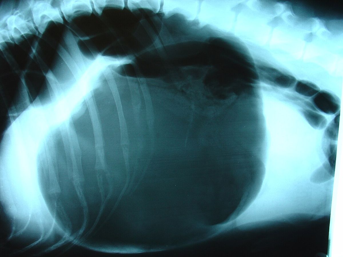
The preferred radiographic view is right lateral recumbency, which shows a large gas-filled stomach with the pylorus displaced dorsally (Figure 2).4 Lactate above 6 mmol/L is linked to a higher chance of gastric necrosis.4 Ventricular arrhythmias are common after surgery. Merck lists heart rate over 140 beats/min, hypotension, poor perfusion, R on T and multifocal premature beats as reasons to start lidocaine.4
Oxygen supplementation
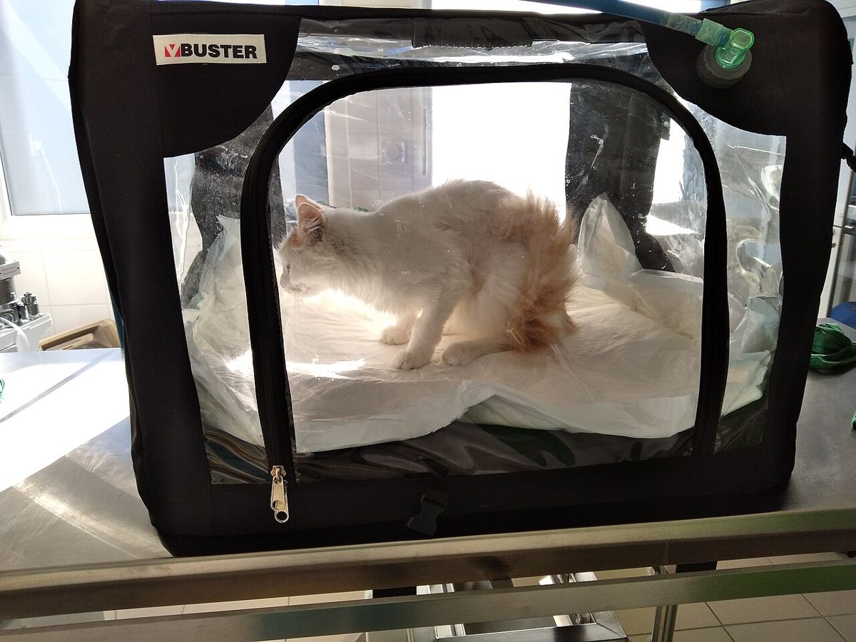
A patient in respiratory distress needs oxygen before restraint, radiographs or blood draws. The method depends on what the patient tolerates and how much inspired oxygen (FiO2, the fraction of inspired oxygen) it needs. Room air is 21% oxygen.
| Method | Flow | FiO2 estimate | Technician notes |
|---|---|---|---|
| Flow-by | 2 to 3 L/min | 25 to 40% | Simple and quick; oxygen line held within a few centimeters of the nostrils |
| Face mask | 1 to 6 L/min | 35 to 55% | FiO2 depends on fit; rebreathing at low flow; ongoing use in conscious patients is limited by poor compliance |
| Oxygen cage | Set by the unit | 21 to 60% | Well tolerated, hands off; FiO2 drops fast when the door opens |
| Nasal catheter or cannula | 50 to 150 mL/kg/min | 30 to 60% | Good for ongoing support; 100 mL/kg/min bilaterally gave a mean FiO2 of 56% and was well tolerated in dogs |
| Oxygen collar (ventral 50 to 75% of an E-collar covered with plastic wrap, tubing taped along the ventral collar) | Varies | Up to about 80% | Can reach a very high FiO2; light sedation may be needed |
| High-flow nasal oxygen | 10 to 60 L/min | 21 to 100% | Heated and humidified; specific equipment; for hypoxemia despite conventional oxygen therapy |
Source: Whitney and Keir 2023 (Table 1 and text); Merck Veterinary Manual, Heart Failure in Dogs and Cats (oxygen collar).56
Seizures and status epilepticus
Status epilepticus must be stopped quickly to prevent death from hyperthermia, acidosis, poor perfusion and hypoxia, and to prevent permanent brain damage.7 Diazepam acts fast and is usually the first drug. An IV bolus can be repeated up to 3 times at 5 to 10 minute intervals.7 Without IV access, diazepam can be given rectally, or midazolam intranasally, as ordered.73 In cats, Reed lists levetiracetam (30 to 60 mg/kg IV) and phenobarbital as later steps.3
Reed recommends oxygen support. Avoid jugular occlusion and a head-down position, because both can raise intracranial pressure.3 Never put fingers or anything else in a seizing animal's mouth. RECOVER advises against giving anything by mouth during active seizures.9
Diabetic ketoacidosis
DKA is decompensated diabetes. Without enough insulin, cells burn fat and the liver makes ketoacids. The result is dehydration, metabolic acidosis, electrolyte losses, vomiting and anorexia.8 Treatment has four parts: IV fluids, regular (short-acting) insulin, potassium and phosphorus supplementation, and care for any underlying disease.8 In two studies, only 38 to 50% of cats with DKA had already been diagnosed with diabetes.3
Diagnosis needs hyperglycemia, ketonemia and acidosis. Urine dipsticks measure acetoacetate. A handheld ketone meter measures beta-hydroxybutyrate.3 Reed notes that euglycemic DKA can occur in cats given SGLT2 inhibitor drugs, so a normal glucose does not rule out DKA in those cats.3
Heat stroke
Heat stroke is a core temperature above 41°C (105.8°F in dogs) with central nervous system dysfunction.9 It can follow a hot environment or exercise, even on a cool day. Exertional heat stroke is reported in dogs but not in cats.9 The 2026 RECOVER first aid guidelines recommend moving the animal somewhere cool and cooling it actively with cool running water on the trunk, especially the thinly haired ventral abdomen, soaking to the skin. Cooling paws alone is not recommended.9 Stop active cooling and dry the animal at 39.7 to 40°C (103.5 to 104.0°F), then transport it immediately.9
Dystocia
Dystocia means difficult birth. Merck lists these warning signs: strong contractions for more than 30 minutes without a neonate; more than 2 hours between neonates without contractions; active or weak labor over 4 hours with no neonate; total whelping or queening over 24 hours; and dark green or foul discharge before the first birth, which can mean placental separation.10 Normal fetal heart rate is 200 to 250 beats/min.10 Oxytocin (2 to 5 U per animal, at 20 to 30 minute intervals, no more than 3 doses) is used only when imaging shows no obstruction. If calcium gluconate is ordered, it is given slowly IV about 10 minutes before oxytocin, never SC or IM, with heart rate and rhythm monitored.10
Proptosis
Proptosis is a sudden forward displacement of the globe, with the eyelids trapped behind it. It usually follows blunt trauma, and brachycephalic breeds are predisposed.11 The exposed cornea dries and ulcerates, so the first job is to keep it moist with lubricant. The veterinarian replaces the globe under general anesthesia and places a temporary tarsorrhaphy, which usually stays 14 to 21 days.11 About 25% of dogs recover vision. The outlook for vision in cats is grave.11
Critical care monitoring sheets
The Rule of 20 is a list of 20 parameters that should be checked at least daily in every critically ill animal, many of them several times a day.12 A good flow sheet turns this into a schedule: times down the side, parameters across the top, and the ordered frequency for each. Perfusion is judged from heart rate, membrane color, pulse quality and mentation, plus measured values such as blood pressure, urine output and lactate.12 Write down the actual number, the time, and your initials, and flag values outside the limits the veterinarian set.
Procedure or technique
Technician steps for a blocked male cat, from arrival to the ward:123
- Triage on arrival. Record heart rate, rectal temperature, respiratory rate, mentation and bladder size. Tell the veterinarian at once if the heart rate is under 120 beats/min or the cat is cold.
- Attach the ECG and keep it running. Look for the hyperkalemic pattern in Figure 1.
- Place an IV catheter and collect blood for electrolytes, renal values, acid-base status and PCV, as ordered.
- Start crystalloid fluids and give calcium gluconate, dextrose or insulin exactly as ordered, slowly, watching the ECG.
- If the cat is too unstable for heavy sedation, prepare for decompressive cystocentesis. Merck describes it as generally safe.
- Prepare for urethral catheterization under sedation or general anesthesia, with analgesia. Monitor blood pressure, heart rate and respiratory rate throughout.
- Connect the urinary catheter to a closed collection system. Measure urine output every 4 hours and report output above 2 mL/kg/h.
- Expect the veterinarian to recalculate fluids every 4 hours to match urine output plus maintenance. Check glucose every 4 hours if insulin and dextrose were given.
Flowchart
Species differences
- Cats: urethral obstruction is mainly a male cat emergency.1 Cushing's reflex (high blood pressure with bradycardia from raised intracranial pressure) is seen less often in cats than in dogs.3 The intranasal midazolam route has not been evaluated in cats.3 Vision after proptosis is rarely saved in cats.11
- Dogs: GDV is mainly a large and giant breed disease.4 In obstructed dogs, passing a urinary catheter may relieve the blockage simply, and the pelvic urethra can be felt on rectal examination.1 Proptosis is most common in brachycephalic breeds.11
- Heat stroke: exertional heat stroke is reported in dogs, not cats. Cats get environmental heat stroke, for example when shut in a car or clothes dryer.9
- Large animals: diazepam is also routinely used for emergency seizure treatment in large animal species.7
Calculation
Worked example: calcium gluconate for a blocked cat. A 5 kg cat has tented T waves and absent P waves. The veterinarian orders 10% calcium gluconate at 1 mL/kg slowly IV over 15 minutes. The Merck range is 0.5 to 1.5 mL/kg.1
Volume = 5 kg × 1 mL/kg = 5 mL (the kg cancels, leaving mL).
Rate over 15 minutes = 5 mL ÷ 15 min = 0.33 mL/min, or about 20 mL/h on a syringe pump (0.33 mL/min × 60 min/h).
Check: the whole Merck range for this cat is 2.5 to 7.5 mL (5 kg × 0.5 to 1.5 mL/kg). Keep the ECG running for the whole infusion.
Second example: nasal oxygen. A 20 kg dog is ordered 100 mL/kg/min by nasal catheter. Flow = 20 kg × 100 mL/kg/min = 2,000 mL/min = 2 L/min.5
Safety, legal and client education
- Never give calcium gluconate SC or IM, because it can cause tissue necrosis.10
- Diazepam and midazolam are DEA Schedule IV controlled substances.14 Log every dose drawn up and given in the controlled drug record.
- Tell clients not to predraw diazepam into plastic syringes for home use, because the drug sticks to the plastic and is lost.7 Rectal diazepam for home use in dogs with cluster seizures can be given up to 3 times in 24 hours.7
- Heat stroke advice for phone triage: cool first with running water on the trunk, then come in. Do not attempt cooling if it puts the person at risk.9
- Owners of blocked cats should know that straining in the litter box can look like constipation but is an emergency in a male cat.1
- Oxygen supports combustion. Keep flames and sparks away from oxygen cages and tubing.
Common mistakes and exam traps
- Thinking calcium gluconate lowers potassium. It protects the heart; dextrose, insulin and relieving the obstruction lower potassium.1
- Reading a slow heart rate in a blocked cat as reassuring. Slow and cold points to severe hyperkalemia.3
- Choosing the left lateral view for GDV. The preferred views are right lateral and dorsoventral.4
- Cooling a heat stroke patient to normal temperature. Stop at 39.7 to 40°C (103.5 to 104.0°F).9
- Trusting a negative urine dipstick in DKA. Dipsticks measure acetoacetate, not beta-hydroxybutyrate.3
- Calling flow-by the best oxygen method. It is quick but gives a low, variable FiO2 and is least preferred for ongoing therapy.56
Memory aids
Slow and cold, potassium is bold: a blocked cat with heart rate under 120 and hypothermia probably has potassium above 8 mmol/L (memory aid).
Calcium shields, sugar shifts: calcium gluconate protects the heart; dextrose with insulin shifts potassium into cells (memory aid).
Tall T, lost P, wide QRS: the three ECG signs of hyperkalemia to spot in lead II (memory aid).
Five minutes makes status: a seizure over 5 minutes is status epilepticus (memory aid).
Stop at 104: stop active cooling of a heat stroke patient at about 104°F (40°C) (memory aid).
30, 2, 4, 24: dystocia warning times: 30 minutes of strong straining, 2 hours between neonates, 4 hours of labor without a neonate, 24 hours in total (memory aid).
Quick review
- Male cats are predisposed to urethral obstruction, mostly from urethral plugs.
- Bradycardia under 120 beats/min plus hypothermia in a blocked cat is 98% specific for potassium above 8 mmol/L.
- Hyperkalemia ECG: small or absent P waves, long PR, wide QRS, tall tented T waves.
- Calcium gluconate 10%, 0.5 to 1.5 mL/kg slowly IV, protects the heart but does not lower potassium.
- After unblocking, urine output target is 1 to 2 mL/kg/h; above 2 suggests postobstructive diuresis.
- GDV: large-bore cranial vein catheters, fluids and decompression together; right lateral radiograph.
- GDV lactate above 6 mmol/L is linked to gastric necrosis.
- Flow-by gives about 25 to 40% FiO2; a nasal catheter gives about 30 to 60%; an oxygen cage loses oxygen when opened.
- Status epilepticus: seizure over 5 minutes or repeated seizures without recovery; diazepam IV up to 3 times.
- DKA treatment: fluids, regular insulin, potassium and phosphorus, and the underlying cause.
- Heat stroke: cool with running water on the trunk to 39.7 to 40°C, then stop, dry and transport.
- Proptosis: keep the cornea moist; about 25% of dogs regain vision.