Introduction
Shock is the hemodynamic and cellular change that follows a failure of normal circulation, so that oxygen and fuel delivery to the tissues no longer meets demand.1 Left untreated, it ends in organ failure and death. Early recognition of the type and stage of shock, followed by timely fluid therapy, prevents or reduces that injury.1
The technician is usually the person who places the catheter, runs the bolus, and measures the patient again a few minutes later. This guide covers the four shock types, how shock looks in dogs and in cats, the perfusion parameters you measure, the bolus and reassess approach, hypertonic saline, colloids, resuscitation end points and lactate.
Key facts
- Shock is classed as hypovolemic, cardiogenic, distributive or obstructive, and more than one type is often present.1
- In dogs, persistent tachycardia is often the first and only sign of early (compensatory) shock.4
- Cats rarely show the compensatory stage. A cat in shock is often cold, slow and hypotensive: gray membranes, weak pulses, hypothermia and a normal or low heart rate.46
- Isotonic crystalloids are given as boluses of 10 to 15 mL/kg (low volume) or 20 to 30 mL/kg (high volume) over 10 to 15 minutes, repeated until end points are reached.4
- For cats, Merck gives a smaller initial crystalloid dose of 5 to 10 mL/kg, and small-volume techniques are recommended in hypovolemic cats.2
- The minimum blood pressure goal is a mean arterial pressure above 60 mm Hg (systolic above 90 mm Hg).5
- Normal blood lactate is less than 2 mmol/L, and resolving a high lactate is linked to better survival.75
- Hypertonic saline (7%) at 4 mL/kg IV must not be used in dehydrated patients or patients at risk of hypernatremia.4
How the VTNE tests this
- Recognize shock and its stage from a set of vital signs (Domain 8 Task B: triage of shock).11
- Calculate a bolus volume and an infusion rate from body weight and mL/kg (Domain 8 Task D: fluid resuscitation; Domain 1 Task C).11
- Choose what to measure after a bolus, and what result means the patient responded (Domain 8 Task E: ongoing evaluation).11
- Identify species traps, above all the cold, bradycardic cat, and fluids that are wrong for a given patient. Knowledge areas K13 and K35.
Core concepts
The four types of shock
Normal circulation needs a working heart, enough blood volume, and blood vessels with normal tone and an open path. A problem in any one of these sets off the sympathetic nervous system, which tries to keep tissues perfused.1 Table 1 sorts the four types. Hypovolemia plays a part in every type, which is why so many shocked patients respond to fluids alone.1
| Type | What fails | Example causes | Treatment priority (veterinarian directs) |
|---|---|---|---|
| Hypovolemic | Blood volume deficit of 15% or more | Hemorrhage, severe vomiting and diarrhea | Fluid boluses; stop bleeding; blood products for hemorrhage |
| Cardiogenic | The heart fails as a pump | Cardiomyopathy, valve insufficiency, arrhythmias, tamponade | Fluids can harm; drugs such as antiarrhythmics and inotropes |
| Distributive | Blood is pooled by peripheral vasodilation | Anaphylaxis, sepsis and systemic inflammation, hypoadrenocorticism | Fluids first; vasopressors may be needed |
| Obstructive | Blood flow is physically blocked | Pericardial effusion, emboli, gastric dilatation and volvulus (GDV) | Relieve the obstruction, for example pericardiocentesis |
Source: Merck Veterinary Manual, Fluid Therapy in Animals, and Reed 2026.16
Compensated and decompensated shock
Merck describes three stages of hypovolemic shock in dogs.4
- Early compensatory stage: tachycardia, pink to red membranes, rapid capillary refill time (CRT), bounding pulses. The dog is usually alert. Pain and anxiety can look the same, so analgesia and time to settle help you tell them apart.
- Middle (early decompensatory) stage: pale membranes, prolonged CRT, weak pulses, tachycardia, and reduced responsiveness.
- Terminal (late decompensatory) stage: bradycardia, severe hypotension, absent pulses, gray or ashen membranes, prolonged CRT, depressed mentation, low urine output and abnormal breathing.
Decompensation happens when the body's compensating responses are used up and cells can no longer make enough energy (ATP).2
Perfusion parameters
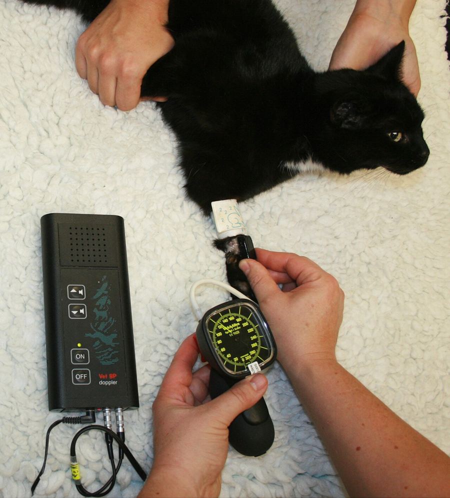
Perfusion means blood flow that delivers oxygen to tissues. The sympathetic response shows up as changes in heart rate, pulse intensity, blood pressure, CRT, membrane color, level of consciousness and rectal temperature. These physical parameters, plus blood pressure, are how the team detects a blood volume deficit.2 Measured values such as blood pressure, urine output and blood lactate add objective data.5 Figure 1 shows them together.
Lactate
When tissues are poorly perfused or short of oxygen, cells make lactate, and blood lactate rises (type A lactic acidosis).5 Normal lactate is less than 2 mmol/L.7 One high value matters less than the trend: resolving hyperlactatemia after fluid resuscitation is associated with better survival to discharge.5 In cats in shock, lactate is typically above 2.5 mmol/L, but it is not specific for shock.6
Fluid choices
Isotonic crystalloids (for example lactated Ringer's solution) are the first-line resuscitation fluid. Crystalloids replace interstitial volume, while colloids act mainly as intravascular volume replacement.2
Hypertonic saline (3%, 7% or 7.5%) rapidly draws water from the interstitial space into the blood vessels.2 Merck gives 7% at 4 mL/kg IV, and notes possible benefit in traumatic brain injury.4 It is best used when hydration is normal and together with other fluids. In a dehydrated patient, or one with free water loss such as heatstroke or diabetes, it can cause severe hyperosmolality with neurologic signs.2
Colloids are fluids with molecules too large to pass freely across the capillary wall. Natural colloids include plasma, albumin and whole blood; synthetic colloids include hydroxyethyl starch (HES).2 Given with crystalloids (HES 2 to 10 mL/kg IV bolus), they reduce the crystalloid volume needed.4 Synthetic colloids are linked to acute kidney injury and coagulation problems. Risk seems low at small doses (under 20 mL/kg) or short use (under 24 hours), and they are not advised in patients that are already azotemic.2 For hemorrhage, blood products are often the ideal choice.2
Procedure or technique
The 2024 AAHA Fluid Therapy Guidelines move away from one-size-fits-all fluid rates. They call for careful initial assessment, judicious boluses and frequent reassessment, and warn against giving too much fluid.8 The technician's part of a bolus looks like this:
- Record a baseline. Heart rate, pulse quality, membrane color, CRT, mentation, rectal temperature, blood pressure and, if ordered, lactate and PCV/TS. You cannot judge a response without a starting point.
- Get IV access fast. Place a short, wide catheter as directed. In tiny or hypotensive patients, especially pediatric ones, an intraosseous catheter can be placed in the femur or humerus after aseptic preparation.6
- Confirm the order. Fluid type, mL/kg, total mL and time (for example 15 mL/kg over 15 minutes). Calculate the volume and check it with a second person.
- Give the bolus by pump, pressure bag or syringe so it actually runs in the ordered time.
- Warm the patient during resuscitation. Use warm air blowers, warm water blankets and IV fluid warmers; avoid hot water bottles and electric blankets, which can burn.42
- Reassess right after each bolus. Repeat the baseline measurements and report the trend to the veterinarian.2
- Watch for fluid overload: faster or harder breathing, crackles, serous nasal discharge, chemosis (swelling of the conjunctiva), jugular distension, shivering, edema or hypertension.10 Stop and tell the veterinarian.
| Fluid | Typical bolus (veterinarian orders) | Watch for |
|---|---|---|
| Isotonic crystalloid, dog | 10 to 15 mL/kg (low volume) or 20 to 30 mL/kg (high volume) over 10 to 15 min | Response after each bolus; overload signs |
| Isotonic crystalloid, cat | 5 to 10 mL/kg (Merck); Reed uses 10 to 15 mL/kg over 15 min | Hypothermia, pulmonary edema, heart disease |
| Hypertonic saline 7% | 4 mL/kg IV (dog); 7.5% at 2 to 4 mL/kg over 10 min once for head trauma in cats | Dehydration, hypernatremia risk |
| Hydroxyethyl starch | 2 to 10 mL/kg IV bolus with crystalloids | Kidney injury, bleeding; avoid if azotemic |
| Blood products | Per transfusion order | Transfusion reactions |
Source: Merck Veterinary Manual (Initial Triage; Fluid Resuscitation Plan) and Reed 2026.426
Flowchart
Figure 2 shows the resuscitation and reassessment loop. Every arrow returns you to measuring the patient again.
Species differences
Dogs show the classic hyperdynamic picture early: fast heart rate, bounding pulses, rapid CRT.4
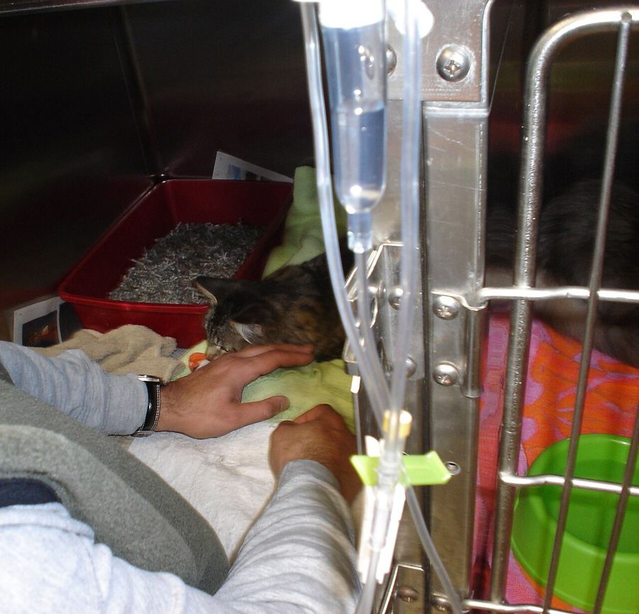
Cats often skip that stage. Instead they show bradycardia (below 160 bpm), hypothermia (below 37.8 C; 100 F) and hypotension (systolic below 90 mm Hg), with dull mentation, pale membranes and slow CRT.6 Hypothermia, especially in cats, blunts the response to the body's own catecholamines and to fluids. Giving large volumes to a cold cat without active warming can cause pulmonary edema while the cat is still hypotensive.2 That is why cats get smaller boluses and are warmed at the same time. Reed advises reassessing heart rate, blood pressure and mentation after every bolus. A response usually appears after 20 to 30 mL/kg, and if there is little response at 30 mL/kg, more fluid is unlikely to help.6 Rule out heart disease first: fluids can harm a cat in cardiogenic shock.6
Small-volume techniques are also recommended for any dog with closed-cavity hemorrhage, head injury, lung contusions or edema, cardiogenic shock, or oliguric kidney failure.2
Calculation
Worked example. The veterinarian orders a 15 mL/kg bolus of lactated Ringer's solution over 15 minutes for a 22 kg dog. What volume, and what pump rate in mL/h?
Volume: 22 kg x 15 mL/kg = 330 mL (kg cancels, leaving mL).
Rate: 330 mL / 15 min x 60 min/h = 1,320 mL/h (min cancels, leaving mL/h).
Cat check: a 4 kg cat ordered 5 mL/kg receives 4 x 5 = 20 mL. Over 15 minutes that is 20 / 15 x 60 = 80 mL/h.
Old method, for comparison: older texts started with one quarter of a "shock dose" of 90 mL/kg for dogs and 44 mL/kg for cats.9 For the 22 kg dog that is 0.25 x 90 x 22 = 495 mL. Current guidance doses each bolus by mL/kg and reassesses after each one.48
Safety, legal and client education
- The veterinarian prescribes fluid type, volume and rate. The technician gives the order, measures the response and reports; do not change a resuscitation plan on your own.
- Label every bag that has an additive and every line, so hypertonic saline is never run by mistake as maintenance fluid.
- Warming devices can burn a patient that cannot move. Check skin under the device and never use electric blankets or hot water bottles.2
- When talking with owners, explain that shock is a whole-body circulation emergency, that the first hour is spent stabilizing before tests are complete, and that the team rechecks the patient after every dose.
Common mistakes and exam traps
- The "normal" heart rate cat. A cat with a heart rate of 150 bpm, low temperature and gray gums is in shock, not stable.
- The full shock dose. Answers that give 90 mL/kg to a dog in one go reflect outdated teaching. Pick small boluses with reassessment.
- Hypertonic saline in a dehydrated patient. It pulls water out of tissues that are already dry. Wrong choice.
- Fluids for every shock. In cardiogenic shock, extra fluid can worsen congestion. Check for heart disease first.
- One lactate value. The trend after treatment tells you more than a single result.
- Pain mimics compensated shock in dogs; treat pain and let the patient settle before calling it shock.4
Memory aids
"Has Cardio Damaged Oxygen?": Hypovolemic, Cardiogenic, Distributive, Obstructive, the four shock types (memory aid).
Cold, slow, low: the shocked cat is hypothermic, bradycardic and hypotensive (memory aid).
Bolus, then look: every bolus is followed by a full reassessment of heart rate, pulses, membranes, CRT, mentation and blood pressure before the next one (memory aid).
60, 2, 1: MAP above 60 mm Hg, lactate below 2 mmol/L, urine output at least 1 mL/kg/h (memory aid).
Salt needs water: hypertonic saline pulls water from the interstitium, so it needs a hydrated patient (memory aid).
Quick review
- Shock types: hypovolemic, cardiogenic, distributive, obstructive.
- Hypovolemic shock follows a blood volume loss of 15% or more.
- Early canine shock: tachycardia, red gums, fast CRT, bounding pulses.
- Late shock: bradycardia, gray gums, absent pulses, depressed mentation.
- Cats in shock are often bradycardic, hypothermic and hypotensive.
- Dog crystalloid bolus: 10 to 15 or 20 to 30 mL/kg over 10 to 15 minutes.
- Cats get smaller boluses with active warming.
- Reassess after every bolus.
- Minimum targets: MAP above 60 mm Hg, systolic above 90 mm Hg.
- Normal lactate is below 2 mmol/L; a falling lactate is a good sign.
- No hypertonic saline in dehydrated or hypernatremia-risk patients.
- Synthetic colloids carry kidney and bleeding risks; avoid in azotemic patients.