Introduction
Every anesthetic carries risk, and most problems show up on the monitor or in your hands before they become disasters. The 2020 AAHA guidelines list the common complications as hypotension, hypoventilation, hypoxemia, hypothermia and some arrhythmias such as sinus tachycardia and bradycardia, and they call cardiovascular and respiratory problems the most acutely life-threatening.1 As the anesthetist, the technician spots the change, checks the patient and the equipment, starts the first steps the practice protocol allows and tells the veterinarian. This guide covers each common complication, what you see, the likely cause and your response, plus regurgitation, rough or slow recoveries, equipment faults and the crash cart.
Key facts
- AAHA hypotension: systolic below 80 to 90 mm Hg, mean below 60 to 70 mm Hg, or diastolic below 40 mm Hg. The AAFP cat guidelines use systolic below 90 and mean below 60.13
- Hypoxemia is SpO2 below 95%; severe hypoxemia is below 90%. Cyanosis appears only when hypoxemia is profound.1
- Start positive pressure ventilation (PPV) when EtCO2 is above 60 mm Hg, after ruling out excessive depth.1
- Hypothermia is a core temperature below 98 °F (36.7 °C). It slows drug metabolism and prolongs recovery.1
- Bradycardia rules of thumb: below 60 beats/min in dogs and below 25 in adult horses; in cats a heart rate below 100 may reduce cardiac output.23
- Regurgitation under anesthesia: suction the esophagus, then lavage with saline or tap water while the cuffed tube protects the airway.1
- Between 47% (dogs) and 60% (cats) of anesthetic-related deaths happen after the procedure, most within the first 3 hours.1
How the VTNE tests this
- Anesthesia is 20 of the 150 scored items (13%). Task F asks you to "monitor and respond to patient status during all stages of anesthesia (pre-, peri-, and post-)".6
- Knowledge statement K42 covers monitoring, recovery and troubleshooting; K43 covers drug reactions and side effects.6
- Expect scenario items: a number on the monitor, then "what should the technician do first?"
- Expect threshold recall (SpO2, MAP, EtCO2, temperature, heart rate) and simple dose or bolus volume calculations.
- Expect equipment questions: closed pop-off valve, exhausted absorbent, failed leak test.
Core concepts
Hypotension means blood pressure too low to perfuse the brain, heart, kidneys and other organs. Lee lists two main causes: anesthetic overdose and hypovolemia from bleeding or a fluid deficit.2 Inhalants cause dose-dependent hypotension, so reducing the inhalant is usually the first step.1
Hypoventilation means too little air moves in and out, so carbon dioxide builds up (hypercapnia). It causes respiratory acidosis and can cause hypoxemia. Capnography measures it; watching chest movement is subjective. AAHA gives EtCO2 of about 35 to 45 mm Hg awake and 40 to 50 (up to 55) mm Hg at a surgical plane.1 Apnea is no breathing at all. After induction many patients are apneic or breathe slowly, and AAHA advises giving 1 to 4 breaths/min by hand until breathing returns.1
Hypoxemia means low oxygen in arterial blood. It is uncommon when an intubated patient breathes 100% oxygen, so a low SpO2 sends you looking for a cause: a misplaced or disconnected tube, an empty oxygen supply, hypoventilation, tube placement past the thoracic inlet into one bronchus (one-lung intubation), collapsed lung (atelectasis) in obese or bloated patients on their backs, lung or pleural disease, or poor perfusion.1
Hypothermia is the most predictable complication. AAHA lists delayed drug metabolism, cardiovascular problems, poor perfusion, breathing problems, cerebral depression and more wound infections.1 Very small dogs and all cats are at extra risk.1
Bradycardia (slow heart rate) comes from deep anesthesia, high vagal tone (brachycephalic breeds, opioids) and parasympathetic stimulation such as a distended bladder, handling of the viscera or the oculocardiac reflex. Tachycardia comes from light anesthesia, drugs such as atropine, reflex response to hypotension and hypercapnia. Premature ventricular contractions (PVCs) usually follow acidemia, hypoxia, hypercapnia or pain, and can progress to ventricular fibrillation.2 AAHA also says tachycardia can come from hypoxemia or hypovolemia, so do not assume it means the patient is too light.1
| Complication | What you see | Likely causes | Technician response |
|---|---|---|---|
| Hypotension | MAP below 60 to 70, weak pulses, slow CRT | Too deep, hypovolemia, bleeding | Check depth, reduce inhalant, tell the DVM, fluid bolus or drugs as ordered |
| Hypoventilation | EtCO2 above 60, shallow or slow breaths | Too deep, drugs, rebreathing CO2 | Check depth and vaporizer, give PPV, check absorbent and valves |
| Apnea | No breaths, flat capnogram | Induction drugs, too deep, arrest | Confirm tube and pulse, give 1 to 4 breaths/min, reduce inhalant |
| Hypoxemia | SpO2 below 95%, late cyanosis | Tube or O2 problem, hypoventilation, atelectasis, poor perfusion | Work through the hypoxemia tree (Figure 2) |
| Hypothermia | Temperature below 98 °F, slow recovery | Heat loss, cold fluids, wet coat | Warm air or warm-water blanket, warm fluids, insulate feet |
| Bradycardia | Dog below 60, cat below 100 | Too deep, vagal tone, opioids, viscera traction | Check depth and surgery, tell the DVM, anticholinergic as ordered |
| Regurgitation | Fluid at mouth or nose, tube or esophagus | Gastroesophageal reflux | Keep cuff up, suction, lavage esophagus |
| Delayed recovery | Not alert well beyond 30 min | Too deep, hypothermia, hypoglycemia | Warm, check glucose, ask about reversal |
Source: 2020 AAHA Anesthesia and Monitoring Guidelines;1 Lee, Anesthetic Emergencies and Accidents;2 AAFP Feline Anesthesia Guidelines.3
Procedure: responding to a complication
- Confirm the reading. Look at the patient: pulse quality, mucous membranes, chest movement, depth signs. AAHA warns that low SpO2 tempts people to fiddle with the probe; check the patient first.1
- Check depth. Read the vaporizer setting and the depth signs. Excessive depth causes hypotension, hypoventilation and bradycardia.12
- Check the airway and machine. Tube in place and connected, oxygen flowing, pop-off valve open, absorbent not exhausted.1
- Tell the veterinarian what you see, with numbers and the trend, and what you have checked.
- Start the treatment ordered or allowed by protocol: reduce inhalant, fluid bolus, PPV, warming, anticholinergic.
- Recheck and record. Note the event, the drug and dose, and the patient's response on the anesthetic record.
Hypotension. Reduce the inhalant, and add opioids or other injectable drugs to lower the inhalant need. AAHA describes an IV crystalloid bolus of 5 to 20 mL/kg and/or a colloid bolus of 1 to 5 mL/kg. If the patient is also bradycardic, an anticholinergic (atropine, glycopyrrolate) or ephedrine is given; if contractility is poor, a positive inotrope such as dopamine or dobutamine is used.12
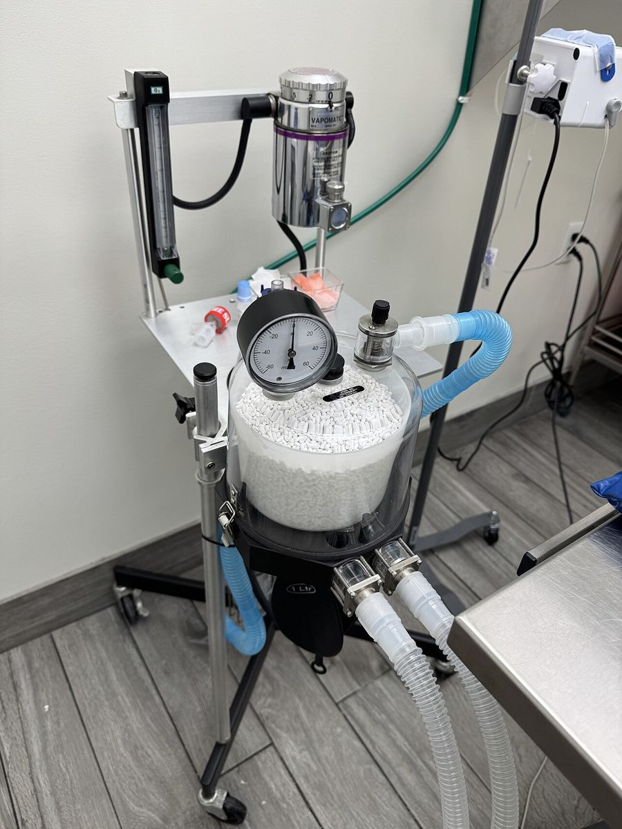
Hypoventilation. First make sure the patient is not too deep. Then give breaths by squeezing the reservoir bag while the adjustable pressure limiting (APL, pop-off) valve is closed, and open it again after each breath. If PPV lowers blood pressure, reduce peak airway pressure. If CO2 stays high, look for excess dead space, exhausted absorbent, sticking one-way valves in a rebreathing circuit or too little oxygen flow in a non-rebreathing circuit. If the machine is faulty, swap machines.1
Hypothermia. Circulating warm-water blankets and warm-air systems work best. Warm IV fluids, a fluid line warmer and insulation on the feet slow heat loss. Never use heat sources not made for anesthetized patients, because they can cause severe burns. Give oxygen to a shivering patient, because shivering raises oxygen use.1
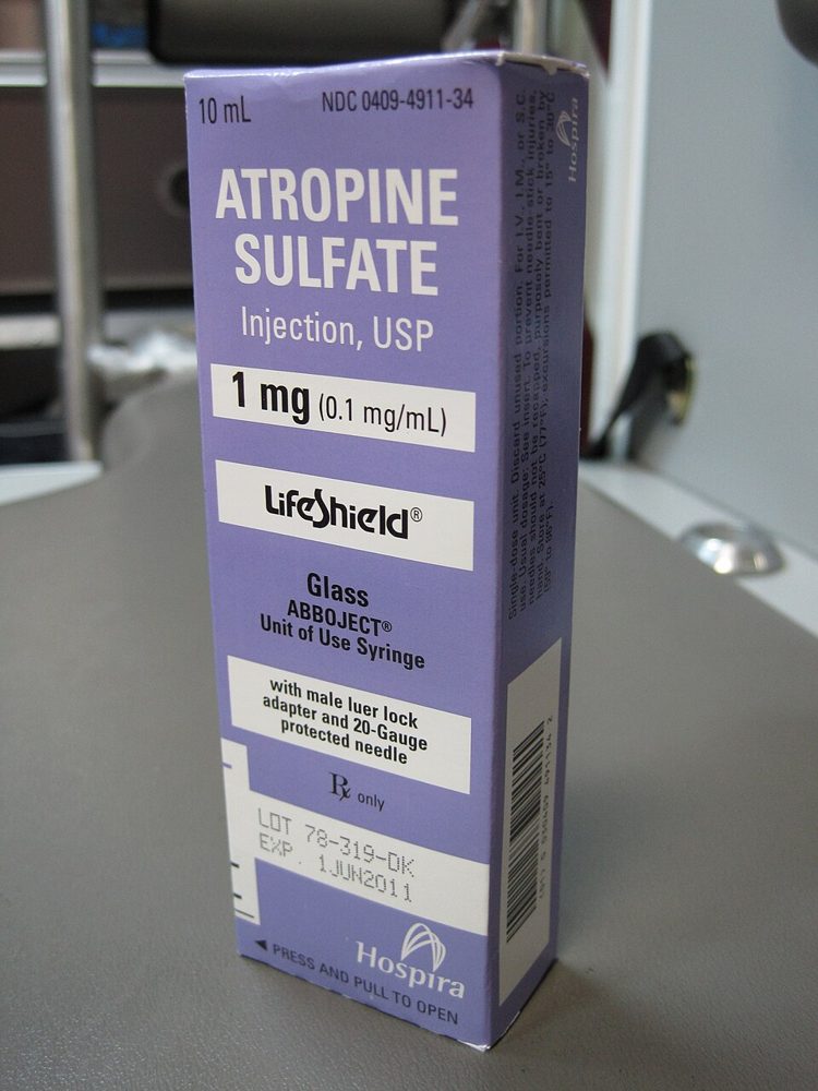
Bradycardia. Look for the cause first: depth, a vagal stimulus from surgery, or drugs. Lee gives atropine 0.02 to 0.04 mg/kg IV and glycopyrrolate 0.005 to 0.01 mg/kg IV for dogs and cats; the veterinarian decides the drug and dose.2 Lidocaine is the usual first drug for PVCs that need treatment, after the cause is corrected.2
Regurgitation. Gastroesophageal reflux (GER) is stomach contents flowing back into the esophagus; regurgitation is when it reaches the mouth. It can cause esophagitis, aspiration pneumonia and, in extreme cases, esophageal stricture. Keep the cuff inflated, suction the esophagus, then lavage it with saline or tap water. Diluted bicarbonate may be instilled to raise esophageal pH.1 Note it on the record so the patient is watched for aspiration.
Flowcharts: hypotension and hypoxemia
Recovery problems and equipment faults
Delayed recovery. AAHA gives an optimal recovery time of 10 to 30 minutes after the end of anesthesia for dogs and cats. Slow recoveries come from too much depth during maintenance, hypothermia and, in small or neonatal patients, hypoglycemia, so check blood glucose. Careful reversal may be considered if recovery is very prolonged.1
Emergence delirium versus dysphoria. Emergence delirium is uncontrolled, uncoordinated thrashing as the patient regains partial consciousness soon after the anesthetic is stopped. Dysphoria has several causes, including uncontrolled pain and hypoxemia from airway obstruction. Check the airway and give oxygen if breathing is the problem. Give an analgesic if pain is suspected; if pain control is adequate, an anxiolytic sedative such as dexmedetomidine (0.001 to 0.005 mg/kg IV or IM) may be ordered.1
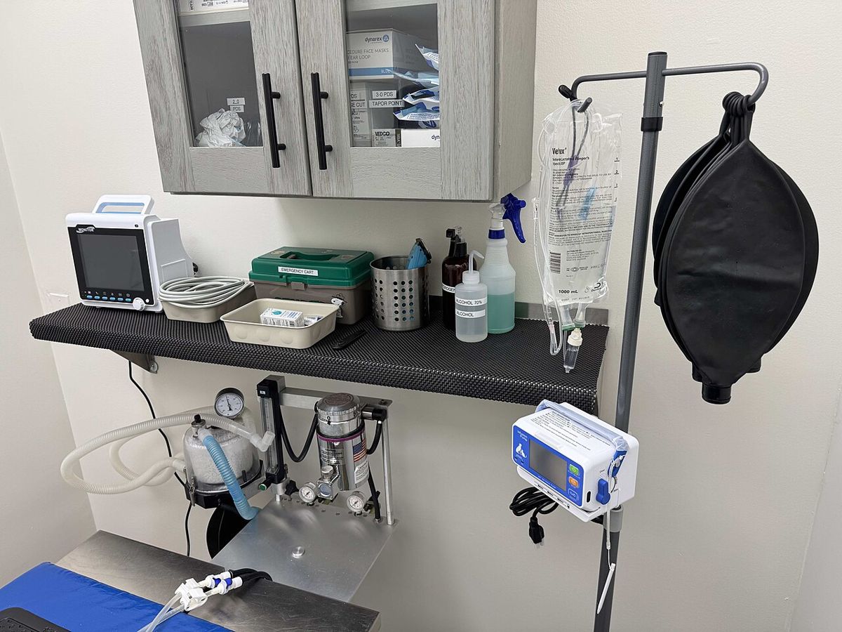
Equipment faults. AAHA calls an in-circuit manometer and a safety pop-off valve the two essential safety features of every machine. A closed pop-off valve lets airway pressure climb quickly and risks barotrauma (lung injury from pressure).1 Lee lists human errors: a decimal slip that gives ten times the dose, mislabeled syringes, the wrong agent in a vaporizer and a hypoxic gas mixture.2 Perivascular injection of an irritant drug causes pain and tissue damage; Lee advises giving drugs through a catheter and diluting a leak with saline.2
Cardiac arrest and the crash cart. Lee's signs of arrest are no pulse or heartbeat, apnea, loss of corneal reflexes, fixed wide-open eyes and dilated pupils that do not respond to light.2 Merck notes that arrest linked to anesthesia has a better prognosis than other arrests. RECOVER recommends standardized, regularly audited resuscitation equipment, including a crash cart with suction and a defibrillator, plus posted dose charts and CPR algorithms.5 The CPR guide covers the full sequence.
| Finding | Why it is urgent | While you wait |
|---|---|---|
| No pulse, no heartbeat or apnea with dilated pupils | Possible cardiac arrest | Call the code, turn off inhalant, bring the crash cart |
| SpO2 below 90% | Severe hypoxemia | Check tube and O2, ventilate |
| MAP still low after reducing inhalant | Organs underperfused | Recheck BP, have fluids ready |
| New PVCs or an irregular rhythm | May progress to ventricular fibrillation | Print or save the ECG strip, check SpO2 and EtCO2 |
| Fluid at the mouth or nose | Aspiration risk | Keep cuff inflated, ready suction |
Source: 2020 AAHA Anesthesia and Monitoring Guidelines;1 Lee, Anesthetic Emergencies and Accidents;2 Merck Veterinary Manual, CPR.5
Species differences
- Dogs. Bradycardia is below 60 beats/min as a general rule.2 Brachycephalic dogs have narrow airways and high vagal tone. Lee advises pre-oxygenation, avoiding drugs that cause vomiting such as xylazine and morphine, leaving the tube in as long as possible and watching for upper airway obstruction in recovery.2
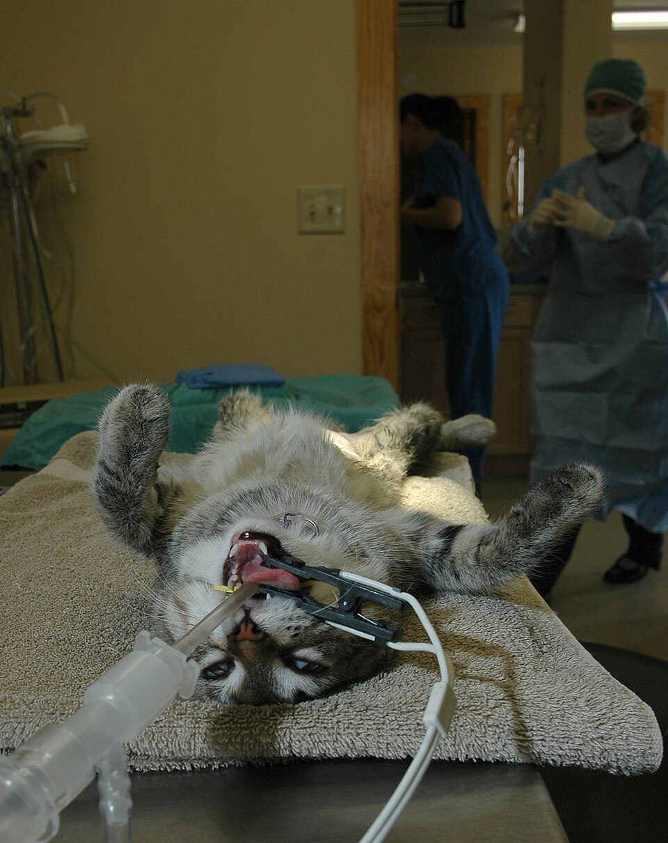
- Cats. A heart rate below 100 may reduce cardiac output, and hypotension is common in cats on inhalants when there is no surgical stimulation.3 Most feline anesthetic deaths happen within 3 hours after anesthesia and are commonly caused by airway obstruction. Overinflated cuffs can tear the trachea.3
- Horses. Bradycardia is below 25 beats/min in adults.2 Lee advises keeping MAP above 60 mm Hg, or 70 mm Hg in heavily muscled breeds, and padding and positioning the limbs to prevent post-anesthetic myopathy, which may appear 1 to 2 hours after the horse stands.4

Calculation
Worked example. A 20 kg dog is hypotensive and bradycardic. The veterinarian orders glycopyrrolate 0.01 mg/kg IV and a crystalloid bolus of 10 mL/kg. Glycopyrrolate is supplied at 0.2 mg/mL.7
Glycopyrrolate dose: 20 kg x 0.01 mg/kg = 0.2 mg (kg cancels).
Volume: 0.2 mg / 0.2 mg/mL = 1.0 mL (mg cancels).
Bolus: 20 kg x 10 mL/kg = 200 mL (kg cancels). This sits inside the AAHA range of 5 to 20 mL/kg.1
Safety, legal and client education
- Treatments are given on the veterinarian's order or a written practice protocol. Record every event, drug, dose, route and time on the anesthetic record.
- Keep emergency drugs, a dose chart for the patient's weight and suction ready before induction. AAFP recommends calculating patient-specific emergency doses and volumes in advance.3
- Monitor recovery with the same vigilance as maintenance, because most anesthetic deaths happen in the first 3 hours after the procedure.1
- Tell clients that a patient may seem fine in the hospital but show lethargy, nausea or vomiting at home, and ask about previous anesthetic recoveries at admission.1
Common mistakes and exam traps
- Trusting mucous membrane colour. Cyanosis appears only when hypoxemia is already profound; use pulse oximetry.1
- Assuming tachycardia means "too light". It can also mean pain, hypoxemia, hypercarbia or hypovolemia.1
- Leaving the pop-off valve closed after a manual breath. Pressure builds and can cause barotrauma.1
- Turning the vaporizer up when the patient moves, without checking blood pressure first. Inhalant hypotension is dose dependent.1
- Using an ordinary heating pad. Use devices designed for anesthetized patients to avoid burns.1
- Deflating the cuff when the patient regurgitates. Keep it up to protect the airway while you suction.1
Memory aids
60, 60, 60: a dog heart rate below 60, a MAP below 60 and an EtCO2 above 60 all need action (memory aid). Sources: Lee for heart rate, AAHA for MAP and PPV.
95 and 90: SpO2 below 95% is hypoxemia; below 90% is severe (memory aid, AAHA thresholds).
Cat 100: in an anesthetized cat, a heart rate below 100 may already lower cardiac output (memory aid, AAFP).
Tube, Tank, Breath, Blood: the hypoxemia check order: tube placed and connected, oxygen supply, ventilation, perfusion (memory aid, AAHA steps).
Cuff up, suck out, wash out: regurgitation response: keep the cuff inflated, suction the esophagus, lavage with saline or tap water (memory aid, AAHA).
Quick review
- The common complications are hypotension, hypoventilation, hypoxemia, hypothermia and some arrhythmias.
- Hypotension: systolic below 80 to 90, MAP below 60 to 70, diastolic below 40 mm Hg.
- First response to hypotension: check depth and reduce the inhalant.
- Crystalloid bolus 5 to 20 mL/kg or colloid 1 to 5 mL/kg, as ordered.
- EtCO2 above 60 mm Hg: give PPV after checking depth.
- Hypoxemia is SpO2 below 95%; check tube, oxygen, ventilation, then perfusion.
- Hypothermia is below 98 °F (36.7 °C); use warm air or warm-water blankets.
- Bradycardia: dog below 60, cat below 100, adult horse below 25.
- Regurgitation: keep the cuff up, suction and lavage the esophagus.
- Recovery should take about 10 to 30 minutes; check temperature and glucose if slow.
- Emergence delirium is thrashing at partial consciousness; rule out pain and hypoxemia first.
- Every machine needs a manometer and a safety pop-off valve.