Introduction
Three monitors answer three different questions during anesthesia. The capnograph asks: is the patient breathing out carbon dioxide (CO2) normally? The pulse oximeter asks: is the hemoglobin carrying oxygen? The blood pressure monitor asks: is there enough pressure to push blood through the organs? The veterinary technician sets up these monitors, reads the numbers and the waveforms, fixes equipment problems and tells the veterinarian when a value moves out of range.
This guide covers the four phases of a normal capnogram, causes of high and low end-tidal CO2, rebreathing, esophageal intubation, how pulse oximetry works and when it misleads, and how Doppler, oscillometric and direct arterial blood pressure compare. Depth of anesthesia and the anesthetic record are covered in the anesthetic monitoring guide.
Key facts
- AAHA: EtCO2 is about 35 to 45 mm Hg in awake patients and about 40 to 50 (up to 55) mm Hg at an appropriate surgical plane. Start positive pressure ventilation (PPV) if EtCO2 is above 60 mm Hg.3
- The inspiratory baseline of the capnogram should be zero. Inspired CO2 above 5 mm Hg means the patient is rebreathing CO2.12
- An EtCO2 reading near 0 after intubation suggests the tube is in the esophagus, because very little CO2 is present there.1
- A sudden fall in EtCO2 can be an early sign of cardiopulmonary arrest. A sudden rise during CPR can mean the heart has started beating on its own.1
- Pulse oximetry measures hemoglobin saturation, not ventilation. A shallow-breathing cat on oxygen can show a normal SpO2 while EtCO2 is high.1
- AAHA: SpO2 below 95% is hypoxemia; below 90% is severe. An SpO2 of 90% matches an arterial oxygen tension (PaO2) of about 60 mm Hg.23
- A mean arterial pressure (MAP) below 60 mm Hg means organ and tissue perfusion is inadequate.2
- Cuff width should be about 40% of the limb or tail circumference for Doppler and oscillometric readings.4
How the VTNE tests this
- Read a capnogram: name the shape (normal, rebreathing, obstruction, leak, apnea, arrest) and pick the first action. Domain 7 Task F, "Monitor and respond to patient status during all stages of anesthesia"; K42 includes anesthesia troubleshooting.7
- Interpret a number: is this EtCO2, SpO2 or blood pressure acceptable, and what does it mean?
- Spot the artifact: a low SpO2 from a bad probe site, motion or poor perfusion rather than true hypoxemia.
- Choose the blood pressure method for a small cat, a hypotensive patient or a long critical case.
- Calculate: MAP from systolic and diastolic pressure; non-rebreathing fresh gas flow from body weight.
Core concepts
Capnography
Capnography is the measurement and graphic recording of CO2 in exhaled gas. CO2 diffuses easily from blood into the alveoli, so in healthy patients the arterial CO2 (PaCO2) and alveolar CO2 are almost the same.1 End-tidal CO2 (EtCO2) is the CO2 level at the end of exhalation. It gives a breath-by-breath estimate of PaCO2 without drawing arterial blood.2 A capnograph shows the waveform (the capnogram), the EtCO2 value and the respiratory rate. A capnometer shows numbers only.2
Ventilation and PaCO2 move in opposite directions. When alveolar ventilation doubles, PaCO2 halves.1 Inhalants and opioids depress breathing, so rising EtCO2 (hypercapnia) is the most common change you will see.1 Watching the chest only shows that breathing is present, not whether it is adequate.1
A normal capnogram has four phases.2
- Phase I, inspiratory baseline: fresh gas passes the sensor. It should read zero; if not, the patient is rebreathing CO2.
- Phase II, expiratory upstroke: CO2 reaches the sensor as exhalation begins. Usually very steep.
- Phase III, expiratory plateau: alveolar gas. Its peak is the EtCO2.
- Phase IV, inspiratory downstroke: the next breath starts and CO2 falls steeply to zero.
Shippy describes the normal shape as a top hat or square.1
Mainstream and sidestream capnographs
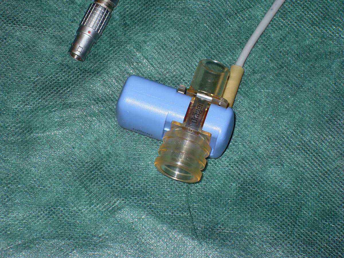
A mainstream sensor sits on the endotracheal tube and the patient breathes through it, so the reading is almost real time. It is heavier and adds dead space. A sidestream unit pulls a gas sample through tubing to the monitor. Expect a delay of 2 to 3 seconds. The line can clog with secretions or water, and high oxygen flow can dilute the sample and give a falsely low reading.1 Dead space is any part of the circuit where gas flows both ways, between the patient's nose and the Y-piece.1
Pulse oximetry
A pulse oximeter detects pulsing arterial blood in a tissue bed, calculates the percentage of hemoglobin carrying oxygen (SpO2) and shows the pulse rate.2 It shines red (660 nm) and infrared (940 nm) light through tissue; oxygenated and deoxygenated hemoglobin absorb these wavelengths differently.5 An anesthetized animal breathing oxygen should read 99 to 100%.2 Because a patient on 100% oxygen is usually protected from hypoxemia, a reading below 90% means the patient is in serious trouble. The AAFP calls the pulse oximeter a crisis monitor and recommends a unit that shows a waveform.4 Arterial blood gas analysis is the gold standard for oxygenation; PaO2 on 100% oxygen should be above 200 mm Hg.2
Blood pressure
Blood pressure shows whether blood flow to the tissues is adequate. Normal values in anesthetized dogs and cats are systolic 90 to 120 mm Hg and diastolic 55 to 90 mm Hg. MAP = diastolic + (systolic minus diastolic) / 3.2 AAHA defines anesthetic hypotension as systolic below 80 to 90 mm Hg, mean below 60 to 70 mm Hg, or diastolic below 40 mm Hg.3 The AAFP feline guidelines use systolic below 90 and MAP below 60 mm Hg.4
| Parameter | Target under anesthesia | Act when |
|---|---|---|
| EtCO2 | 40 to 50 (up to 55) mm Hg at a surgical plane | Above 60 mm Hg: PPV. Below 35 mm Hg: look for the cause. |
| Inspired CO2 | 0 mm Hg | Above 5 mm Hg: rebreathing. |
| SpO2 on oxygen | 99 to 100% | Below 95%: hypoxemia. Below 90%: severe. |
| Systolic pressure | 90 to 120 mm Hg | Below 80 to 90 mm Hg: tell the veterinarian. |
| MAP | 60 mm Hg or more | Below 60 mm Hg: perfusion inadequate. |
Sources: AAHA 20203; Shippy 20241; Lee2.
Procedure: setting up and reading the three monitors
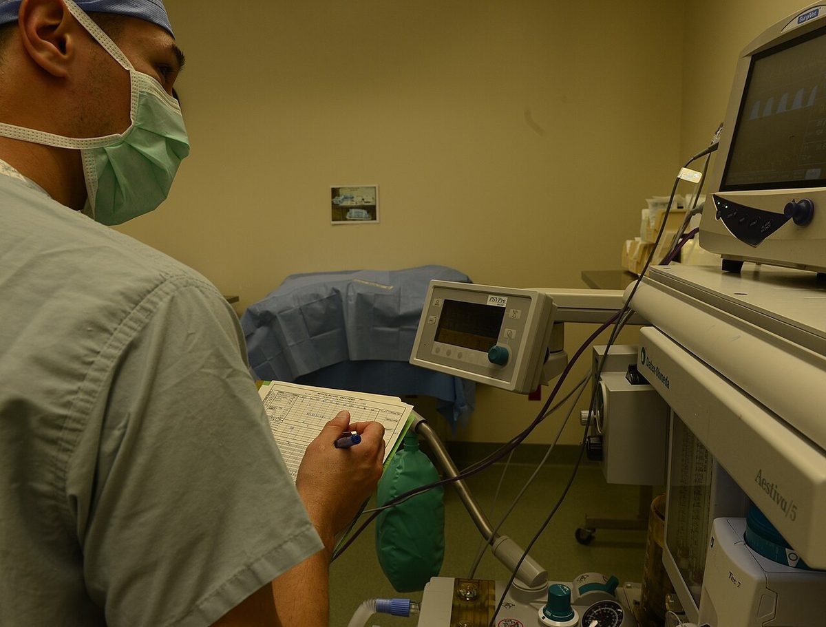
- Capnograph on at intubation. Place the adapter on the end of the tube during intubation. A clear EtCO2 tracing is one way AAHA lists to confirm the tube is in the trachea, along with seeing the tube pass between the arytenoids, bag movement and condensation in the tube.3 No CO2 after a breath points to esophageal placement.1
- Keep dead space small. Elbow and capnograph adapters, long tubes and spirometry sensors all add dead space. Use a pediatric adapter in a small patient.13
- Place the pulse oximeter probe on the tongue, ear, lip folds, toe pads, axillary or inguinal skin fold, prepuce or vulva.2 Check that the pulse rate on the oximeter matches the heart rate.
- Choose and size the blood pressure cuff. Measure the limb or tail and pick a cuff about 40% of that circumference. Place it on the most cylindrical part of the limb.4
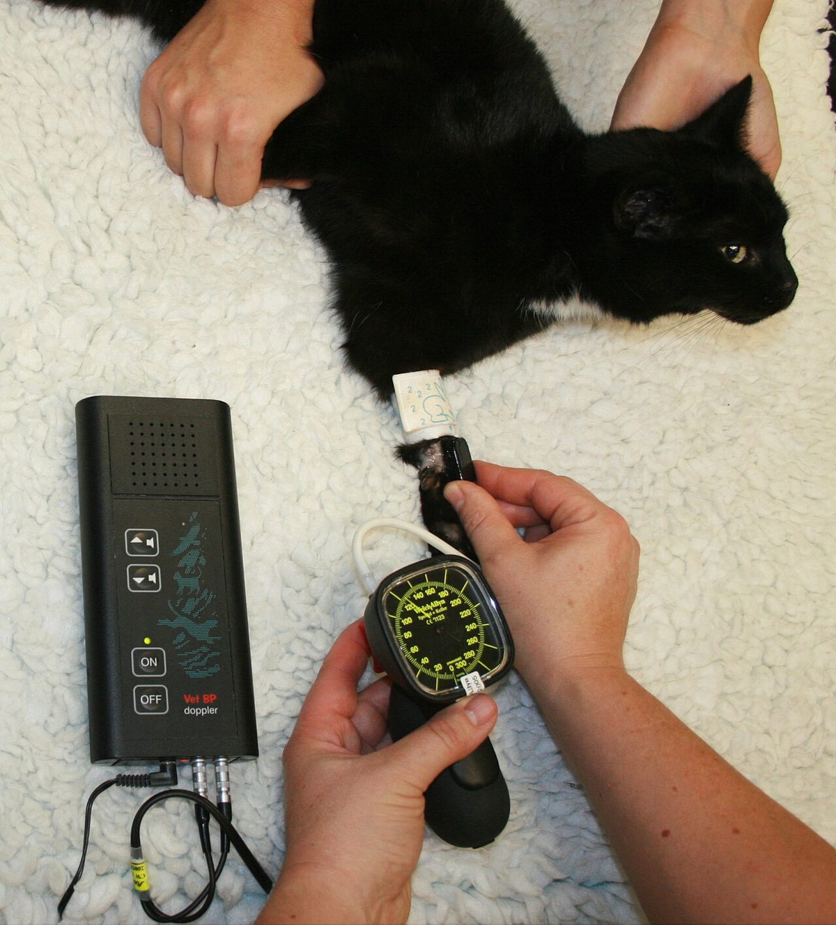
- Doppler technique. Place the probe over an artery: the superficial palmar arterial arch (forelimb), the dorsal pedal artery (hind limb) or the ventral tail base. Inflate the cuff, then read the pressure as the cuff pressure is reduced.8 The Doppler gives systolic pressure and an audible pulse. Deflating the cuff too quickly causes errors.4
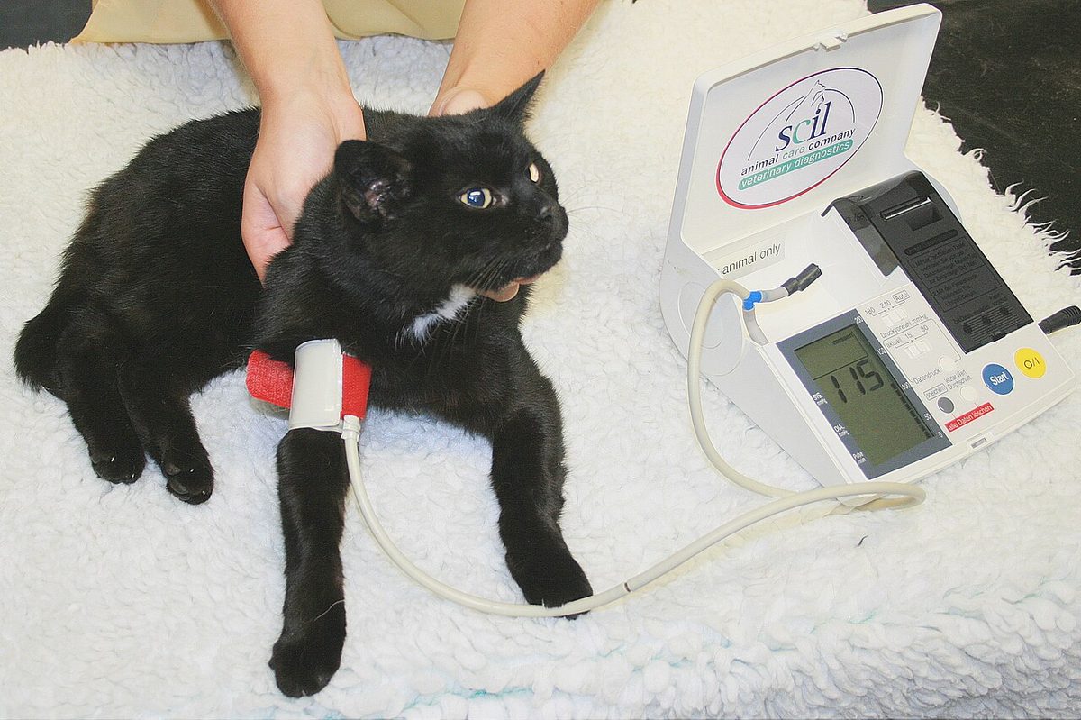
- Oscillometric technique. Set the cycle interval. Lee sets 3 to 5 minute intervals.2 Oscillometric accuracy falls in hypotensive patients and in small patients under 5 kg, while the Doppler method works in animals of any size.2
- Read trends, not single numbers. Record all values on the anesthetic record. A gradual rise in EtCO2 means something different from a sudden drop.
- Look at the patient before the screen. When a value changes, check breathing, pulse and mucous membranes, then the equipment.
Reading the capnogram
| Waveform or value | Likely cause | Technician action |
|---|---|---|
| Normal shape, EtCO2 rising slowly above 45 to 60 mm Hg | Hypoventilation from opioids, inhalants, obesity or endocrine disease | Check depth and the vaporizer; tell the veterinarian; give breaths by bag or ventilator if above 60 mm Hg |
| Baseline does not return to 0 (inspired CO2 above 5 mm Hg) | Rebreathing. Circle: exhausted absorbent, stuck expiratory valve, extra dead space. Non-rebreathing: fresh gas flow too low | Change absorbent, check valves, remove dead space; on a non-rebreathing circuit raise the flow until the baseline is 0 |
| Sloped upstroke ("shark fin") | Obstruction: bronchoconstriction or secretions in the tube | Check the tube for kinks or mucus; tell the veterinarian |
| Back of the square missing | Leak, often at the cuff | Check cuff inflation and connections |
| Abrupt loss of waveform | Extubation, disconnection or apnea | Check the patient and tube; reconnect; ventilate |
| Sudden fall, then flat | Cardiopulmonary arrest, or pulmonary thromboembolism | Check pulse and heartbeat now; call the veterinarian; start CPR if arrested |
| Gradual fall below 35 mm Hg | Hyperventilation (light plane, pain, fever, hypoxemia), sample dilution, more alveolar dead space | Check depth, pain, temperature and SpO2 |
Sources: Shippy 20241; AAHA 20203.
In a rebreathing (circle) system, lower flows are acceptable because the absorbent removes CO2. Condensation can make the lightweight expiratory valve stick.1 In a non-rebreathing system, fresh gas flow does the washing out. AAHA gives about 200 to 400 mL/kg/min for non-rebreathing circuits and says to adjust flow with the capnograph so inspired CO2 stays below 5 mm Hg.3 Shippy gives 200 to 300 mL/kg/min.1
In healthy patients, mild to moderate hypercapnia (50 to 55 mm Hg) can be tolerated and may support hemodynamic stability. Above 70 mm Hg, CO2 acts as a depressant.1 If PPV is excessive, EtCO2 falls below the level that stimulates breathing, so ventilator settings are adjusted based on EtCO2.3
Pulse oximeter pitfalls
| Problem | Effect on reading | What to do |
|---|---|---|
| Motion, including shivering | Erratic or no reading | Check the waveform before trusting the number |
| Ambient or fluorescent light; electrocautery noise | Erratic reading | Recheck once the interference stops |
| Hypotension or vasoconstriction | Weak signal, falsely low or no reading | Try another site; check blood pressure |
| Carboxyhemoglobin (smoke inhalation) | Falsely high | Do not trust a normal SpO2; tell the veterinarian |
| Methemoglobin | Pushed toward 85% | Tell the veterinarian; blood gas needed |
Choosing a blood pressure method
| Method | What it gives | Strengths | Limits |
|---|---|---|---|
| Doppler ultrasonic | Systolic pressure; audible pulse rate and rhythm | Any size, from a rat to a horse; inexpensive; reliable in cats | Needs a skilled operator; labor intensive; may underestimate systolic pressure in anesthetized cats |
| Oscillometric | Systolic, diastolic and mean | Automated; set intervals; frees the anesthetist | Less accurate in hypotensive or small (under about 5 kg) patients |
| Direct (invasive) arterial | Continuous beat-to-beat pressure and waveform | Gold standard; most accurate when hypotensive; allows arterial blood samples | Needs an arterial catheter and transducer; technically harder |
Sources: Lee2; AAFP 20184; Chen 20236.
Flowchart: EtCO2 abnormal
Use this tree when the capnograph alarms or the EtCO2 trend changes. It ends in technician actions; the veterinarian decides on drugs and treatment.
Species differences
- Cats and small dogs. Shallow breathing can keep SpO2 normal on oxygen while EtCO2 climbs, so capnography matters most here.1 Oscillometric readings are less reliable under about 5 kg; Doppler is reliable in patients of all sizes.24 Doppler may underestimate systolic pressure in anesthetized cats.6 Use a pediatric capnograph adapter to cut dead space.1
- Dogs. Obese dogs and dogs with hypothyroidism or hyperadrenocorticism may hypoventilate because abdominal fat or large organs push the diaphragm forward. They may need PPV.1
- Horses. EtCO2 underestimates PaCO2 much more in large animals than in small animals, because of atelectasis and ventilation-perfusion mismatch. Use EtCO2 for trends and check at least one arterial blood gas.5 The pulse oximeter clip usually goes on the tongue, which gives the most accurate reading; ears, nostrils, lips, vulva and prepuce are less accurate.5
Calculation
1. Mean arterial pressure. An oscillometric monitor in a dog shows 96/48 mm Hg but no mean. MAP = diastolic + (systolic minus diastolic) / 3.2
MAP = 48 mm Hg + (96 mm Hg minus 48 mm Hg) / 3 = 48 mm Hg + 16 mm Hg = 64 mm Hg. This is just above the 60 mm Hg line, so watch the trend closely.
2. Non-rebreathing fresh gas flow. A 4 kg cat is on a non-rebreathing circuit. AAHA gives 200 to 400 mL/kg/min.3
4 kg × 200 mL/kg/min = 800 mL/min; 4 kg × 400 mL/kg/min = 1,600 mL/min. The kg cancels, leaving mL/min. Set about 0.8 to 1.6 L/min, then use the capnogram: if the baseline rises above 0, increase the flow.13
Safety, legal and client education
- Monitoring decreases the odds of anesthetic death. Use both electronic monitors and hands-on checks.3
- Do not silence alarms by habit. The pulse oximeter is easily upset by movement and lighting, so alarms are often ignored; a unit with a waveform helps you tell a real change from artifact.4
- Change CO2 absorbent on schedule. AAHA advises changing it after 8 hours of use; Shippy suggests weekly if surgery is daily.13
- Record EtCO2, SpO2 and blood pressure with the time. The record is a legal document.
- For clients: you can explain that their pet will have its breathing, oxygen level and blood pressure watched by machines and by a trained person for the whole procedure.
Common mistakes and exam traps
- "SpO2 is 98%, so breathing is fine." False. Pulse oximetry does not measure ventilation; check EtCO2.1
- Calling a raised baseline hypoventilation. A baseline above 0 is rebreathing. Fix the equipment or flow first.1
- Mixing up shark fin and leak. A sloped front edge is obstruction; a missing back edge is a leak.1
- Treating a sudden EtCO2 drop as a sensor fault. Check the pulse first. It may be arrest.1
- Trusting a normal SpO2 after smoke inhalation. Carboxyhemoglobin reads falsely high.2
- Thinking a Doppler gives a mean. Doppler gives systolic pressure only.4
Memory aids
TOP HAT IS HAPPY: a normal capnogram is square like a top hat; a sloped front (shark fin) is obstruction, a missing back is a leak (memory aid).
OFF ZERO, CHECK THE GEAR: a baseline above 0 is rebreathing; check absorbent, valves and dead space on a circle, or flow on a non-rebreathing circuit (memory aid).
90 IS 60: SpO2 90% matches a PaO2 of about 60 mm Hg, the line for hypoxemia (memory aid).
MAP IS DIASTOLIC PLUS A THIRD: MAP = diastolic + one third of (systolic minus diastolic); keep it at 60 mm Hg or more (memory aid).
FORTY AROUND: cuff width is about 40% of the limb circumference (memory aid).
Quick review
- Capnogram phases: I baseline (0), II upstroke, III plateau (peak = EtCO2), IV downstroke.
- EtCO2 at a surgical plane: about 40 to 50, up to 55 mm Hg; awake 35 to 45.
- EtCO2 above 60 mm Hg: start PPV.
- Inspired CO2 above 5 mm Hg means rebreathing.
- Circle rebreathing causes: exhausted absorbent, stuck expiratory valve, dead space.
- Shark fin is obstruction; missing back edge is a leak.
- EtCO2 near 0 after intubation suggests esophageal intubation.
- Sudden EtCO2 fall: check for arrest; sudden rise during CPR suggests return of circulation.
- SpO2 below 95% is hypoxemia; below 90% is severe; 90% is about PaO2 60 mm Hg.
- Carboxyhemoglobin reads falsely high; methemoglobin pulls the reading toward 85%.
- Doppler: systolic only, any size; oscillometric: automated, poor under 5 kg; direct arterial: gold standard.
- MAP below 60 mm Hg means poor perfusion; cuff width about 40% of circumference.