Introduction
A suture pattern is the path the needle and strand take through tissue to hold a wound closed. A knot is what keeps that strand from sliding loose. Veterinary technicians do not usually choose the pattern, but they hear it called, set up the right suture and needle, cut ends to the right length, count the throws on a knot, and later remove skin sutures and explain incision care to the owner.
This guide sorts the common patterns into three families: appositional, inverting and everting. It explains interrupted versus continuous closure, the square, surgeon's and granny knots, and what the technician watches for after surgery.
Key facts
- Appositional patterns line the cut edges up side by side. Optimal healing comes with appositional patterns because the cells are already lined up.11
- Inverting patterns turn the cut edges inward and limit exposed suture. Cushing and Lembert are the classic inverting patterns for hollow organs such as the stomach, bladder and uterus.912
- Vertical and horizontal mattress sutures evert (turn outward) the skin edges and are used to relieve tension.11
- The square knot is the most frequently used and most secure knot. Its second throw goes in the reverse direction of the first.2
- A granny knot comes from incorrect technique and is more likely to slip. Uneven tension or pulling upward turns a square or granny knot into a slipping half hitch.2
- An interrupted suture is usually knotted with 4 to 5 throws. A continuous line gets 5 to 6 throws at the start and 6 to 7 at the end.1
- Simple interrupted sutures are placed about 5 mm apart, with bites 3 to 5 mm from the tissue edge.3
How the VTNE tests this
- Name the pattern from a description of how the needle travels, or pick the pattern family (appositional, inverting, everting). Domain 2 Task D (function as a sterile surgical technician); K20 (wound closure, including suture materials and patterns, staples and tissue adhesives).16
- Match the pattern to the tissue: hollow organ, skin under tension, a cosmetic buried closure. K20.16
- Judge knot security: square versus granny, why a surgeon's throw is used, how many throws a continuous line needs. K20.16
- Explain post-operative incision care to an owner and recognize a problem at a recheck. Domain 2 Task D; K20.16
Core concepts
Interrupted versus continuous
In an interrupted pattern each stitch is a separate piece of suture with its own knot. If one knot fails, the others still hold. In a continuous pattern one strand runs the length of the incision with a knot only at the start and the end.4 The whole line depends on those two knots. That is why the end knots get extra throws.1
Some surgeons prefer interrupted sutures because a continuous line may not let pus drain, and a continuous suture pulled too tight can strangle the wound edges.13 A buried continuous closure with absorbable suture needs no removal visit.13
Three pattern families
- Appositional: edges meet flat, end to end. Simple interrupted, simple continuous, cruciate, Ford interlocking and intradermal patterns. Most skin, subcutaneous and body wall closures use these.11
- Inverting: edges fold inward, away from the surface. They help prevent leakage from a lumen and reduce adhesions to exposed suture.12 Cushing and Lembert.
- Everting: edges turn outward, toward the surface. Vertical and horizontal mattress sutures. They hold tension so an appositional layer can heal underneath.11
| Pattern | Interrupted or continuous | Family | Typical use | Strengths and drawbacks |
|---|---|---|---|---|
| Simple interrupted | Interrupted | Appositional | Skin | One bite each side, knot, cut. Each stitch stands alone, so one failed knot does not open the line; many knots to tie.3 |
| Cruciate (cross mattress) | Interrupted | Appositional | Skin | Two simple passes tied as one, forming an X. More efficient than simple interrupted sutures.511 |
| Simple continuous | Continuous | Appositional | Linea alba | Knots only at each end; whole line depends on two knots.4 |
| Ford interlocking | Continuous | Appositional | Skin, especially large animals | Each bite passes through the previous loop. The most common skin pattern in large animals and neater than simple continuous.611 |
| Intradermal (subcuticular) | Continuous | Appositional | Cosmetic skin closure | Buried knots, bites in the dermis parallel to the incision; no sutures to remove.811 |
| Vertical mattress | Interrupted | Everting | Skin under tension | Far bite 8 to 10 mm out, near bite 4 mm out. Relieves tension; everts edges.7 |
| Horizontal mattress | Interrupted | Everting | Skin under tension | Second bite runs back 6 to 8 mm along the incision; both ends on one side. Relieves tension; everts edges.7 |
| Cushing | Continuous | Inverting | Stomach, bladder, uterus | Bites run parallel to the incision into the submucosa, not the lumen.9 |
| Lembert | Continuous | Inverting | Stomach, bladder, uterus | Bites run perpendicular to the incision through serosa and muscularis.10 |
Source: Veterinary Surgery Online, chapter 4 pattern pages; University of Minnesota Large Animal Surgery Supplemental Notes.
Knots and throws
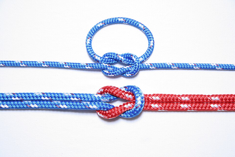
A throw is one wrap of the strand around itself. A knot is two or more throws. The square knot has a second throw in the reverse direction of the first, with even horizontal tension on both ends while it is snugged down. The granny knot has two throws formed the same way, one started with the instrument between the strands and one outside, and it slips more easily. A half hitch forms when tension is uneven, when the strand is pulled upward, or when the second throw goes the same direction as the first.2
A surgeon's knot has an extra pass through the loop of the first throw. The added friction holds the first throw while the second is placed, so it is used in high-tension tissue.2 It is not needed on every stitch.
| Knot or rule | How it is formed | Security and use |
|---|---|---|
| Square knot | Two throws, the second reversed; even horizontal tension | Most used and most secure |
| Surgeon's knot | Extra pass through the loop of the first throw | Holds the first throw in high-tension tissue |
| Granny knot | Two throws started differently, from incorrect technique | More likely to slip than a square knot |
| Half hitch | Uneven tension, upward pull, or same-direction throws | Readily slips; must be followed by square knots if used deliberately |
| Interrupted suture | 4 to 5 throws | Depends on suture material |
| Continuous line | 5 to 6 throws at the start, 6 to 7 at the end | Extra throws for security |
Source: Veterinary Surgery Online, chapter 4: Knots; Ligatures and Suture Patterns.12
Procedure or technique
Assisting with closure (sterile technician)
- Listen for the layer and pattern the surgeon calls, for example "simple continuous on the linea" or "intradermal on the skin". Have the matching suture and needle opened or ready.
- For a subcutaneous or intradermal layer, expect a taper needle for subcutaneous tissue and a cutting needle for the subcuticular layer. Cutting needles are needed for skin in all species.11
- For intradermal closure, have small diameter absorbable suture ready, such as poliglactin 910 (Vicryl) or poliglecaprone 25 (Monocryl) on a cutting needle.8
- Count throws as the surgeon ties. Expect 4 to 5 on an interrupted suture, 5 to 6 at the start of a continuous line and 6 to 7 at its end.1
- Cut ends only where told, with suture scissors, keeping the tips in view so you do not cut the knot or tissue.
- Keep the tissue edges visible with the sponge or retractor so bites can be placed at an even distance from the edge.
Removing skin sutures
- Confirm the removal date and the sutures to remove with the veterinarian's order. Non-absorbable skin sutures and staples are usually removed 10 to 14 days after surgery; buried sutures need no removal.15 Temporary mattress tension sutures may come out after 3 to 4 days, leaving the appositional layer in place.11
- Look before you cut. Note gaps, swelling, discharge or sutures pulled through the skin, and report them. Gaps with sutures missing suggest the patient removed them; a suture present but torn out of one side (cut-out) points to closure under tension or bites placed too close to the edge.14
- Restrain the patient, then lift the knot with thumb forceps and cut one side of the strand close to the skin.
- Pull the suture out by the knot so the part that sat on the surface is not dragged through the tissue.
- Count the sutures removed against the number placed, and record the count and how the incision looked.
Flowchart
Species differences
- Dogs and cats chew and lick at incisions more than large animals.11 Small animals are also less prone to adhesions, so patterns other than inverting ones may be used on hollow organs.12
- Cattle have very thick skin, often closed with size 1 to 3 suture. Horse, pig, sheep and goat skin is closer to dog skin and is closed with 2-0 to 3-0.11
- Large animals: Ford interlocking is the most common skin pattern. Cruciates may be used for non-linear wounds or as a removable drainage suture. Skin staples pinch on removal and are kept for emergencies or non-bovine patients.11
- Ruminants heal quickly, and a wound under heavy tension is often left open rather than forced closed.11
- In large animals, inverting patterns are used to close the intestine, bladder and uterus to prevent leakage and adhesions.12
Calculation
Worked example: estimating how many simple interrupted skin sutures to expect. A 6 cm skin incision will be closed with simple interrupted sutures about 5 mm apart.3
Step 1. Convert length to the same unit: 6 cm x 10 mm/cm = 60 mm.
Step 2. Divide by spacing: 60 mm / 5 mm per suture = 12 sutures (the mm units cancel).
Use the number as an estimate for setup and for checking the count at removal. The surgeon's actual count is the one you record.
Safety, legal and client education
- Handle needles with the needle holder, never fingers, and keep them on the needle counter so the sharps count is correct.
- Tell owners to restrict activity for 7 to 14 days, use a short leash, and prevent jumping and rough play.15
- Do not let the pet lick or scratch the incision; it can pull out sutures and introduce infection. Inspect an unbandaged incision at least twice daily.15
- Do not clean the incision with hydrogen peroxide or alcohol, and do not bathe the pet until the veterinarian allows it.15
- A healthy incision is clean with edges touching and normal or slightly pink skin. Slight redness in the first few days and bruising in pale-skinned dogs can be normal.15
- Common causes of dehiscence (the wound reopening) include closure under tension, sutures too close to the edge, licking and trauma, infection, and premature suture removal.14
Common mistakes and exam traps
- Calling Ford interlocking inverting. It is a continuous appositional pattern; Cushing and Lembert are the inverting pair.6
- Mixing up Cushing and Lembert. Cushing bites run parallel to the incision; Lembert bites run perpendicular to it.910
- Thinking a surgeon's throw belongs on every suture. It is for tissue under high tension.2
- Thinking mattress sutures appose. They evert, and are often temporary tension sutures over an appositional layer.11
- Pulling one strand up while tightening. It turns a square knot into a half hitch that slips.2
Quick review
- Interrupted: a knot on every stitch. Continuous: knots at the two ends only.
- Appositional patterns line edges up and give the best skin healing.
- Cushing (parallel bites) and Lembert (perpendicular bites) invert hollow organs.
- Vertical and horizontal mattress sutures evert and relieve tension.
- Cruciate is two simple passes tied as an X.
- Ford interlocking passes each bite through the previous loop.
- Intradermal closure is buried and uses small absorbable suture on a cutting needle.
- Square knot: second throw reverses the first. Most secure.
- Granny knot slips; uneven or upward pull makes a half hitch.
- Surgeon's knot adds an extra pass on the first throw for high tension.
- Throws: 4 to 5 interrupted; 5 to 6 at the start and 6 to 7 at the end of a continuous line.
- Owners: restrict activity, stop licking, no peroxide or alcohol, check twice daily.