Introduction
Most laboratory errors start before the sample reaches an analyzer. The wrong tube, a half-filled citrate tube, a lipemic sample from a dog that just ate, or a tube of whole blood frozen against an ice pack can all produce numbers that look real and are wrong. Collecting, processing, labelling and shipping samples is technician work, and the VTNE lists it as its own task in the Laboratory Procedures domain.7
This guide covers tube types and additives, the order of draw, how to make serum and plasma, the three common interferences (hemolysis, lipemia and icterus), storage, labelling, chain of custody and how to pack a Category B (UN3373) shipment.
Key facts
- EDTA (purple or lavender top) is the anticoagulant of choice for mammalian CBCs. Some birds, reptiles and fish hemolyze in EDTA, so heparin is used for them.1
- Citrate (blue top) is for coagulation tests. The tube must hold nine parts blood to one part citrate. Underfilling falsely prolongs clotting times.4
- Serum (red top or serum separator tube) is the default chemistry sample. Let it clot 20 to 30 minutes at room temperature before spinning.1
- EDTA contamination of a chemistry sample markedly raises potassium and lowers calcium.2
- Withhold food for 12 hours before chemistry samples when it is safe, because lipemia interferes with many tests.13
- Never freeze whole blood. It lyses the red cells and causes gross hemolysis.1
- Most veterinary diagnostic samples ship as UN3373, Biological substances, Category B, in triple packaging.16
How the VTNE tests this
- Choose the tube for a named test (CBC, chemistry panel, PT and aPTT, glucose). Domain 4 Task B; K24 (sample collection, preparation, analysis, storing and shipping).7
- Put tubes in order of draw and explain why the order matters (additive carryover).
- Predict the artifact: which result goes up or down with hemolysis, lipemia, icterus, EDTA contamination or delayed separation.
- Troubleshoot a rejected or odd sample: underfilled citrate tube, gel serum, a frozen EDTA tube.
- Package and label a sample for an outside laboratory, including the triple packaging layers and what goes on the outer box.
Core concepts
Serum, plasma and whole blood
Serum is the fluid left after blood clots. Plasma is the fluid from blood that was kept from clotting by an anticoagulant. Plasma still contains fibrinogen, so plasma protein reads higher than serum protein.2 Potassium is higher in serum than in plasma because cells release potassium during clotting.2 Hematology needs anticoagulated whole blood, because the cells themselves are counted.1
How the additives work
EDTA and citrate both bind (chelate) calcium, which the clotting cascade needs. Citrate binds it more gently, so the laboratory can add calcium back to run clotting tests.2 Heparin works differently: it boosts the action of antithrombin.2 Every additive tube is made for a set volume of blood. Overfilling or underfilling changes the blood to additive ratio and makes results unreliable.1
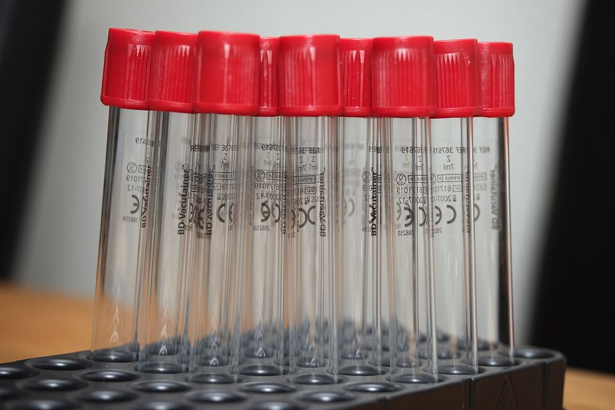
| Cap color | Additive | Main use | Technician notes |
|---|---|---|---|
| Red | None (serum) | Chemistry, serology | Clot 20 to 30 min, rim the clot, spin, then move the serum to a clean tube.1 |
| Striped red ("tiger top") | Separator gel (serum) | Chemistry | Gel sits between cells and serum after spinning. Respin if the gel layer is cracked. Gel is not a perfect barrier.12 |
| Green | Heparin (plasma) | Chemistry, some exotic hematology | Fill more than half full to avoid artifacts such as falsely high bilirubin.2 |
| Purple or lavender | Potassium EDTA | CBC, blood smear, platelet count | Not for potassium, calcium, magnesium, iron or most enzymes.2 |
| Blue | Sodium citrate 3.2% or 3.8% | PT, aPTT and other clotting tests | Fill exactly: 9 parts blood to 1 part citrate.4 |
| Gray | Sodium fluoride and potassium oxalate | Glucose when separation is delayed | Glycolytic inhibitor. Fluoride oxalate can cause hemolysis.53 |
Sources: Merck Veterinary Manual; eClinPath, Cornell University; CLSI.
Order of draw
When several tubes come from one venipuncture, fill them in a set order so that additive carried on the needle cannot contaminate the next tube. The CLSI order is: blood culture, citrate, serum tubes (with or without clot activator or gel), heparin, EDTA, then fluoride oxalate. The order is the same for syringe, tube holder or evacuated tube collection.5 Plastic serum tubes with clot activator can interfere with clotting tests, so only a culture bottle or a tube without activator may go before the citrate tube.5
Interferences: hemolysis, lipemia and icterus
Hemolysis (red or pink serum) is the most common interference. It is usually an in vitro artifact of poor venipuncture, lipemia, frozen whole blood, slow separation or delayed submission.3 Lipemia (milky, white serum) is usually a post-meal artifact. A fast of at least 12 hours prevents most of it, and lipemia in a fasted animal suggests disease.3 Icterus (yellow serum from bilirubin) has little effect on hematology but lowers chemistry creatinine and total protein at high bilirubin levels.3
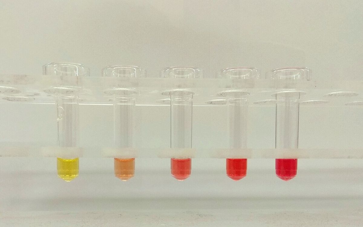
| Interference | Hematology effect | Chemistry effect |
|---|---|---|
| Hemolysis | PCV and RBC count falsely low; MCH and MCHC falsely high; refractometer line blurred; RBC ghosts may be counted as platelets | Falsely high potassium (horses, camelids, some cattle, Akita and Shiba Inu dogs), AST, LDH, magnesium; phosphate rises with storage; CK can rise; water released dilutes other analytes |
| Lipemia | Hemoglobin, MCH and MCHC falsely high; refractometer total protein falsely high; lipid may be counted as platelets | Light scatter can raise some results (bilirubin on some methods); volume displacement lowers sodium and chloride; promotes hemolysis |
| Icterus | Minimal to none, including refractometer protein | Creatinine and total protein decreased at high bilirubin |
Source: eClinPath, Interferences, Cornell University.3 Effects are method dependent; check your analyzer's guidance.
Storage artifacts
Leaving serum or plasma sitting on the cells causes false changes. Cells keep using glucose, so glucose falls. In species with high red cell potassium (horses, Akitas, sheep, some cattle such as Holsteins, camelids) potassium rises. Cold storage slows these changes but does not stop them, and a gel separator tube does not prevent them either.2
Procedure or technique
Making serum
- Confirm the test request and the tube needed. Ask the reference laboratory before collecting for any unusual test or species.1
- Collect into a red-top or serum separator tube.
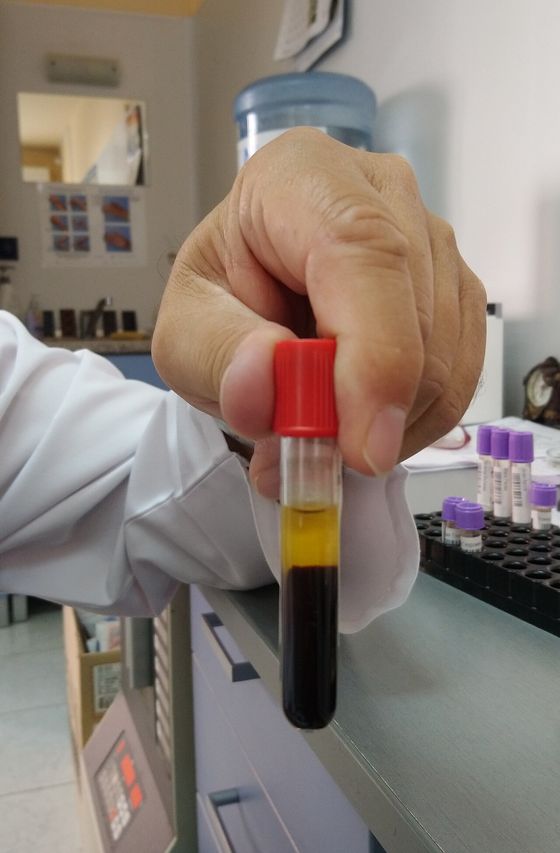
- Stand the tube at room temperature for 20 to 30 minutes so the clot forms and retracts. A clot that is not complete can leave gelled serum from late fibrin.1
- Rim the clot off the tube wall with a wooden applicator stick. Horse and ruminant blood clots slowly and may need rimming more than once.2
- Centrifuge at moderate speed (about 450 g) for 10 minutes.12
- Check a separator tube for a cracked gel layer and respin if needed. Pipette serum from a plain tube into a clean, labelled tube.1
- Refrigerate or freeze the serum until it is run. Merck notes that 0.5 mL of serum is typically preferred for a chemistry panel.1
Hematology samples
- Fill the EDTA tube to its fill volume.1
- Make several blood smears immediately to limit changes in red cell shape, platelet clumping and cell breakdown.1
- Refrigerate the anticoagulated blood. Keep smears at room temperature, away from the cold pack.1
Coagulation samples
- Use a clean, single stick from a large peripheral vein. Tissue contamination activates clotting. If the vein is missed, use a fresh needle; if flow stops partway, start again with a new needle and tube.4
- Fill the citrate tube exactly to the line.4
- Separate the plasma within 6 hours for PT and aPTT, at 1,500 g for 15 minutes, and move it with a plastic pipette into a plastic tube with no additive.1
- Ship plasma chilled to arrive within 24 hours, or freeze it in a dedicated (not frost-free) freezer and ship on dry ice.4
Labelling, records and chain of custody
Label every container with a waterproof marker. At minimum write the patient identification and the specimen type, because urine and serum can look the same.1 The submission form goes with the sample in a waterproof bag. It should carry the owner's name, species, breed or weight, sex, age, animal ID, clinical signs, treatment and relevant earlier results. Flag a suspected zoonotic disease on the form to protect laboratory staff.1 If legal action is possible, seal containers so tampering would show, or hand-carry them and get a receipt, and document the chain of custody.1
Flowchart
Packaging and shipping
Most veterinary diagnostic samples are Category B infectious substances: material not likely to cause permanent disability or life-threatening disease in healthy people or animals on exposure. They ship as "UN3373, Biological substances, Category B." Merck lists two other shipping labels for infectious substances: UN2814 (affecting humans) and UN2900 (affecting animals). If a high-risk or reportable disease is suspected, contact state and federal veterinary authorities before shipping.1
US rules (49 CFR 173.199) require triple packaging for Category B: a leakproof primary receptacle, leakproof secondary packaging with enough absorbent material to soak up the entire liquid contents, and a rigid outer packaging with cushioning.6 The outer box shows the UN3373 diamond (each side at least 50 mm) with the words "Biological substances, Category B" beside it. The name and phone number of a responsible person goes on the box or the shipping document. By air, each primary receptacle holds no more than 1 L and each outer package no more than 4 L of liquid.6
Practical packing points from Merck:1
- Never send needles, scalpel blades, broken slides, syringes or containers without a sealable opening.
- Ship liquids in sealed jars or tubes, not plastic bags.
- Bag cold packs separately and keep them off whole blood, which can freeze and hemolyze. eClinPath suggests wrapping tubes in paper towel.2
- If dry ice is used, mark the box and do not seal the lid with tape.
- Formalin by air is limited to 30 mL of free liquid per inner container and 1 L per package. Fix tissue in house for at least 24 hours, then ship it in a small volume of fresh formalin.
Species differences
- Dog: Akitas and Shiba Inus have high red cell potassium, so hemolysis or delayed separation can falsely raise potassium.23 Fast for 12 hours before chemistry when it is safe; Merck lists pediatric patients and toy breeds as exceptions to consider.1
- Cat: blood volumes are small, so plan the tubes before you stick. Every additive tube must still be filled to its fill volume, or the blood to additive ratio is wrong.1
- Horse and ruminants: blood clots slowly, so rim the clot more than once. Separator tubes are most useful here.2 Horses, sheep, some cattle and camelids have high red cell potassium.2
- Birds, reptiles and fish: some hemolyze in EDTA; heparin is commonly used instead.1
Calculation
Worked example: citrate ratio. You are drawing a coagulation sample into a syringe preloaded with 0.3 mL of 3.2% sodium citrate. How much blood do you draw?
The ratio is 9 parts blood to 1 part citrate.4
Blood volume = 0.3 mL citrate x (9 mL blood / 1 mL citrate) = 2.7 mL blood. The mL citrate units cancel.
Check: total = 2.7 + 0.3 = 3.0 mL, and 0.3 / 3.0 = one tenth citrate. A 5 mL citrate tube works the same way: 0.5 mL citrate and 4.5 mL blood.4
Safety, legal and client education
- Treat every sample as potentially infectious, and mark suspected zoonoses on the form.1
- Package to the federal rule. The shipper must follow the packaging maker's filling and closing instructions.6
- For cases that may go to court (cruelty, poisoning, insurance), keep a documented chain of custody.1
- Client education: explain why the pet should come in fasted for blood work, and when it should not (for example pediatric patients and toy breeds, where the veterinarian may decide against a fast).1
- For paired serology, the first (acute) sample is frozen and the second (convalescent) sample is taken 10 to 14 days later; both go to the lab together.1
Common mistakes and exam traps
- Running potassium or calcium on EDTA plasma, or letting EDTA carry over into a serum tube. Expect high potassium and low calcium.2
- Accepting a half-filled blue-top tube. Underfilling means too much citrate and falsely prolonged clotting times.4
- Reading a high refractometer total protein on lipemic plasma as real. Lipid particles refract light.3
- Leaving serum on the clot overnight. Glucose falls, and potassium can rise.2
- Laying a whole blood tube directly on an ice pack or freezing it. Freezing lyses red cells.1
- Assuming a serum separator tube protects against storage artifacts. It does not; still spin and separate promptly.2
Memory aids
"Cultures Can't Stand Hot Engine Grease" (memory aid): Culture, Citrate, Serum, Heparin, EDTA, Gray fluoride oxalate, the CLSI order of draw.
"Blue is 9 to 1" (memory aid): a citrate tube holds 9 parts blood to 1 part citrate; underfill gives falsely long clotting times.
"Purple pumps potassium" (memory aid): potassium EDTA contamination raises potassium and lowers calcium on chemistry.
"Milky means meal" (memory aid): lipemia is usually post-meal; fast 12 hours, and expect falsely high refractometer protein.
"Three layers, one diamond" (memory aid): Category B is primary, secondary with absorbent, rigid outer, and a UN3373 diamond on the box.
Quick review
- EDTA (purple) for CBC; heparin for some birds, reptiles and fish.
- Citrate (blue) for clotting tests, filled exactly at 9 to 1.
- Serum is the default chemistry sample; clot 20 to 30 minutes first.
- Spin serum at about 450 g for 10 minutes, then separate it.
- Order of draw: culture, citrate, serum, heparin, EDTA, fluoride oxalate.
- EDTA in a chemistry sample: high potassium, low calcium.
- Hemolysis: low PCV, high potassium in some species, high AST and LDH.
- Lipemia: high refractometer protein, high hemoglobin indices; fast 12 hours.
- Icterus: little hematology effect; lowers chemistry creatinine and protein.
- Refrigerate blood, keep smears at room temperature, never freeze whole blood.
- Label patient ID and specimen type with waterproof marker; document chain of custody for legal cases.
- UN3373 Category B: triple packaging, absorbent, rigid outer box, diamond mark.