Introduction
An in-house laboratory is only as good as its equipment and the person running it. Merck notes that in-clinic testing is often done by people with little technical training, so quality control (QC) must be rigorous, and errors can happen in testing, in sample handling and in recording results.10 The veterinary technician keeps the microscope, centrifuge, refractometer and analyzers working, runs and records controls, and keeps the lab safe for people. This guide covers equipment care, calibration, controls, Levey-Jennings charts and basic lab safety.
Key facts
- Total magnification equals ocular power times objective power. A 10X ocular with the 100X oil immersion objective gives 1000X.2
- Immersion oil belongs only on the oil immersion objective. Once oil is on the slide, do not swing the 40X objective through it.2
- Remove immersion oil completely after each slide, using lens tissue.4
- Calibrate an analog refractometer with distilled water: the reading must sit on the 1.000 line, or you turn the calibration screw until it does.5
- Balance the centrifuge, close the lid while it runs, and wait for the rotor to stop completely before opening it. With infectious material, wait 10 minutes after it stops.6
- Control material is usually run daily, and results should fall within the designated range, typically the mean plus or minus 2 standard deviations (SD).7
- On a Levey-Jennings chart, one point outside 3 SD (the 13s rule) rejects the run. One point outside 2 SD (12s) is a warning that triggers a closer look.8
- Employers must keep labels and safety data sheets (SDS) for every hazardous chemical and train staff to handle them.11
How the VTNE tests this
- Maintain equipment: Domain 4 Task D asks you to maintain laboratory equipment and related supplies to ensure safety and quality of results.1
- Read a control chart: decide whether a run is accepted or rejected, and what to do next (K26, quality assurance: maintenance of equipment, verification of test results, calibration and controls).1
- Calculate: total magnification, how many SD a control value is from the mean, or a diluted urine specific gravity.
- Choose the safe action: SDS, PPE, eyewash, sharps and centrifuge safety (K37, environmental health and safety, including hazardous material handling and personal safety).1
Core concepts
The compound microscope
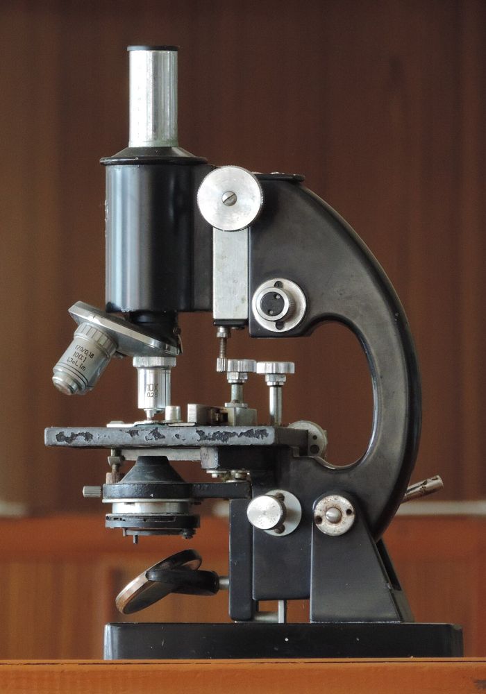
A compound microscope magnifies twice: first through the objective lens, then through the ocular (eyepiece), which usually magnifies 10X. Most have four objectives on a revolving nosepiece: scanning 4X, low power 10X, high dry 40X and oil immersion 100X.2 As magnification goes up, the field of view and the light both go down, so you open the iris diaphragm or turn up the light.2 The condenser, under the stage, concentrates light onto the specimen. Parfocal means a specimen in focus with one objective is nearly in focus with the next, so only the fine adjustment is needed after you change objectives.2
Immersion oil has about the same refractive index as glass, so less light bends away between the slide and the 100X lens. Oil on any other objective gives a fuzzy, distorted image.2
Kohler illumination (Köhler) is a way of setting up the light path, introduced in 1893 by August Köhler, that gives specimen lighting that is uniformly bright and free from glare. Microscope manufacturers recommend it.3 In practice it means centering and focusing the condenser, as in the steps below.
Refractometer
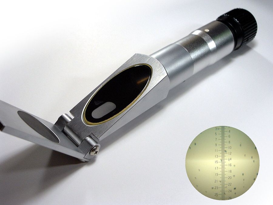
A refractometer compares the refractive index of a sample with distilled water. Urine specific gravity (USG) has no units.5 Common causes of wrong readings include too little sample, calibration error, reading the wrong scale (Brix or protein), failing to close the cover and high temperature.5 Room temperatures outside 16 °C to 38 °C (60 °F to 100 °F) may affect accuracy. Some models are temperature compensated and need calibration less often; always follow the manual.5
Centrifuge
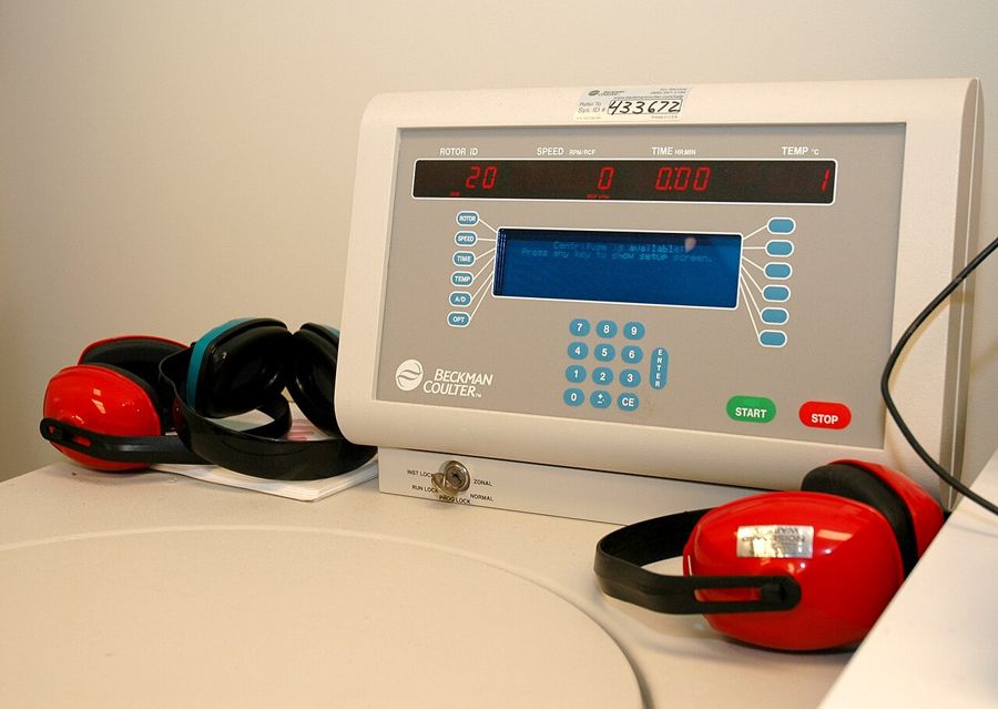
Centrifuges run at high speed and can injure users. OSHA states that unbalanced rotors can cause injury or death, that broken containers release aerosols that are harmful if inhaled, and that most centrifuge accidents are user error.6
Accuracy, precision, calibration and controls
Accuracy is how close a result is to the true value. Precision is how close repeated measurements of the same sample are to each other.7 Bias is a consistent push in one direction; random error makes results jump above and below the mean.7 Running controls means analyzing manufacturer-supplied QC material and comparing the result with the stated mean, range and SD. A control that is out (for example more than 2 SD from the mean) means the instrument may not give accurate results for that test and must be checked.7 Calibration and QC materials are a running cost of in-house testing.10 eClinPath points out that QC is often skipped with point-of-care analyzers, even though the numbers drive treatment decisions.7
Levey-Jennings charts and Westgard rules
A Levey-Jennings chart plots each control result (y-axis) against run or day (x-axis). Lines mark the mean, mean plus and minus 2 SD, and mean plus and minus 3 SD.9 Limits are set from at least 20 measurements over at least 10 days.9 The Westgard multirules use several rules together to keep false rejections low while catching real errors.8
| Rule | What you see | Action |
|---|---|---|
| 12s | One control beyond mean plus or minus 2 SD | Warning: check the other rules |
| 13s | One control beyond mean plus or minus 3 SD | Reject the run |
| 22s | Two consecutive controls beyond the same 2 SD limit | Reject the run |
| R4s | One control above +2 SD and another below -2 SD in the same group | Reject the run |
| 41s | Four consecutive controls beyond the same 1 SD limit | Reject the run |
| 10x | Ten consecutive controls on one side of the mean | Reject the run |
Source: Westgard QC, Westgard Rules (multirule QC).8
Procedure or technique
Microscope: set up, focus and clean
- Carry the microscope with one hand under the base and the other on the arm.2
- Center the condenser: with the 4X objective in place, close the field (base) diaphragm, raise or lower the condenser until its edge is sharp, center it with the centering screws, then open it until the light circle is just larger than the field.2
- Start on 4X. Use the coarse knob to bring the specimen into view, then the fine knob for sharp focus.2
- Move to 10X and 40X with the fine knob only. Never use the coarse knob at high power; it can crack the slide and damage the lens. If you lose focus, go back to 4X.2
- For 100X, turn the nosepiece halfway between 40X and 100X, place a drop of oil on the slide, then rotate the 100X objective into the oil and fine focus.2
- Clean up: rotate to the scanning objective, lower the stage, remove the slide, wipe oil from the 100X lens, clean the lenses, turn down the light and switch off.2 Remove oil with lens tissue rather than solvents, and keep the instrument covered between uses.4
Refractometer: calibrate and read
- Put on gloves. Place a sample-sized drop of distilled water on the prism and close the cover.5
- Hold toward a light source and read. On an analog model, if the line is not on 1.000, turn the calibration screw until it is.5
- Dry the prism, apply the sample, close the cover and read the correct species scale.
- Clean the prism after every use with distilled water and optical tissue or a soft cloth.5
Centrifuge: safe run
- Inspect tubes for cracks; use matched tubes and do not overfill. Make sure the bowl and tubes are dry and the spindle is clean.6
- Balance the load: tubes or containers must be properly balanced in the rotor, so place tubes of equal weight opposite each other.
- Seat the rotor properly, close the lid, and never exceed the rotor's maximum speed.6
- Stay until it runs normally. Open only after a complete stop; wait 10 minutes for infectious material.6
- If a tube breaks, follow the clinic's decontamination procedure and report it to your supervisor.6
Analyzer QC: run, plot, decide
- Run the control material before patient samples, as the analyzer manual directs (usually daily).7
- Compare each result with the target mean and SD from the control insert, and plot it on the Levey-Jennings chart.9
- Apply the rules in Table 1. Record the result, lot number, date and your initials in the QC log.
- If the run is rejected, do not report patient results. Follow Figure 1.
| Equipment | Each use | Each day or on schedule |
|---|---|---|
| Microscope | Remove oil from the 100X objective and slide; clean lenses; cover the instrument | Label oil bottles with the date received and discard old oil4 |
| Refractometer | Clean and dry the prism | Calibrate with distilled water to 1.000; daily is recommended5 |
| Centrifuge | Check tubes for cracks, balance, close lid, wait for full stop | Keep the bowl dry and the spindle clean; service per manufacturer6 |
| Chemistry or hematology analyzer | Check sample quality before loading | Run controls (usually daily) and log them; calibrate and service per manufacturer7 |
Source: FSU Molecular Expressions, Today's Veterinary Nurse, OSHA QuickFacts and eClinPath.4567
Flowchart
Species differences
- Cat versus dog urine: veterinary refractometers include a urine scale calibrated specifically for cats; specific gravity for other species is read on the dog scale.15 Some refractometers read only one species.5
- Analyzer settings: many in-clinic analyzers need the species selected before the run so the right reference interval is printed. Check the species field before you load a horse, ruminant or exotic sample.
- Controls are not species specific: QC material checks the instrument, not the patient, so the same control run covers every species tested that day.7
Calculation
Worked example 1: how far out is the control? A glucose control has a mean of 200 mg/dL and an SD of 4 mg/dL, so the 2 SD limits are 192 and 208 mg/dL.9 Today's result is 211 mg/dL.
SD from mean = (211 mg/dL minus 200 mg/dL) ÷ 4 mg/dL = 11 mg/dL ÷ 4 mg/dL = 2.75 SD (the mg/dL units cancel).
2.75 SD is beyond 2 SD but inside 3 SD: a 12s warning. Check the previous run. If it was also above +2 SD, the 22s rule rejects the run.
Worked example 2: diluted urine. A urine sample reads off the scale. You mix 1 drop urine with 1 drop distilled water; the diluted reading is 1.032. Multiply the digits after the decimal point by 2: 0.032 × 2 = 0.064, so USG is 1.064.5
Worked example 3: magnification. 10X ocular × 40X objective = 400X total.2
Safety, legal and client education
- Hazard communication: OSHA's Hazard Communication Standard, aligned with the Globally Harmonized System (GHS), requires labels and SDS for hazardous chemicals in the workplace and training for the workers who use them.11 An SDS has 16 sections; Section 8 covers exposure controls and personal protection.12
- Eyewash: where eyes or body may be exposed to corrosive materials, OSHA requires facilities for quick drenching or flushing within the work area.13
- No food in the lab: eating, drinking, smoking, applying cosmetics or lip balm and handling contact lenses are prohibited where exposure is reasonably likely.14
- No mouth pipetting of blood or other potentially infectious material.14
- Sharps: do not bend, recap or remove contaminated needles unless no alternative exists, and then only by a one-handed technique or a mechanical device.14
- PPE: wear gloves for samples and reagents and add eye protection for splash risk; the SDS lists the PPE for each chemical.12
- Client education: if an owner asks why a result is delayed, explain plainly that the machine is checked against known samples before results are released, and that a failed check means the test is repeated or sent out so the number can be trusted.
Common mistakes and exam traps
- Using the coarse knob at 40X or 100X. Only the fine knob at high power.2
- Rotating the 40X objective through oil. Go from 100X back to 10X or 4X instead.2
- Calibrating a refractometer with urine or saline. Use distilled water, which reads 1.000.5
- Treating a single 12s point as an automatic rejection in a Westgard question. It is a warning rule; 13s and the others reject.8
- Missing a trend: ten points on one side of the mean (10x) rejects the run even when none is outside 2 SD.8
- Repeating the control until it passes and reporting the patient results without finding the cause.
Memory aids
OIL ONLY ON ONE HUNDRED (memory aid): immersion oil goes only on the 100X objective; never swing 40X through it.
FINE AT FORTY (memory aid): at 40X and 100X use only the fine focus knob.
WATER IS ONE (memory aid): distilled water must read 1.000 on the refractometer.
THREE IS OUT, TWO IS A SHOUT (memory aid): a point beyond 3 SD rejects the run; a single point beyond 2 SD is a warning.
TEN ON ONE SIDE (memory aid): ten consecutive controls on one side of the mean (10x) rejects the run.
STOP, THEN TEN (memory aid): open the centrifuge only after the rotor stops; with infectious samples wait 10 minutes more.
Quick review
- Total magnification is ocular times objective; 10X with 100X is 1000X.
- Objectives: 4X scanning, 10X low, 40X high dry, 100X oil immersion.
- Oil only on the 100X objective; remove it after each slide with lens tissue.
- Fine focus only at high power; parfocal lenses need little refocusing.
- Kohler-style setup centers and focuses the condenser for even, glare-free light.
- Calibrate the refractometer with distilled water to 1.000 and clean the prism after use.
- Balance the centrifuge, close the lid, open only after a full stop.
- Accuracy is closeness to the true value; precision is closeness of repeat results.
- Run controls (usually daily) and log them before reporting patient results.
- 12s warns; 13s, 22s, R4s, 41s and 10x reject.
- A rejected run means hold results, troubleshoot, rerun controls, then call service if still out.
- Know where the SDS are, use the eyewash for corrosive splashes, and never eat or mouth pipette in the lab.