Introduction
Clinical chemistry is the measurement of substances in blood plasma or serum: enzymes, waste products, proteins, glucose and electrolytes. Results help the veterinarian find and monitor disease, and one test is very seldom specific to one condition.1 The veterinary technician's part is to collect the right sample, handle it so it stays true, run the analyzer with quality control and flag anything that could make a number wrong. This guide covers sample types, analyzer types, the common renal, hepatic and metabolic analytes, the three big interferences and the basics of blood gas samples.
Key facts
- Serum comes from clotted blood (red top). Plasma comes from blood with an anticoagulant. Heparin (green top) plasma is the preferred anticoagulant sample for chemistry.4
- Let clotted samples stand at least 30 minutes, then spin and separate serum or plasma from the cells as soon as possible.1
- Cells left in contact with serum use up glucose and can leak potassium, so a delayed sample can read falsely low glucose.4
- EDTA, even a small carry-over, markedly increases potassium and decreases calcium. Never pour purple-top blood into a chemistry tube.4
- Hemolysis, lipemia and icterus are the three common interferences. Hemolysis is the most common, and it is usually caused by collection or handling.5
- Stress can raise blood glucose, especially in cats. Stress uncommonly pushes feline glucose above 288 mg/dL and usually resolves within a few hours.11
- A bile acid panel pairs a fasting sample with a sample taken 2 hours after a meal.7
- Blood gas samples go into a heparinized syringe, are kept free of air and are run promptly at the point of care, not mailed out.9
How the VTNE tests this
- Choose the sample: pick the tube or sample type for a chemistry panel, glucose, potassium or bile acids (Domain 4 Task B).12
- Recognize an artifact: say how hemolysis, lipemia, delayed separation or EDTA contamination changes a result (Domain 4 Task C; K25, K27).12
- Match analyte to organ: link BUN, creatinine and SDMA to the kidney, and ALT, ALP, GGT, bilirubin and bile acids to the liver.
- Calculate: globulin from total protein and albumin, or convert units.
- Run the analyzer: explain quality control and when an in-house result should go to a reference laboratory.
Core concepts
Serum or plasma
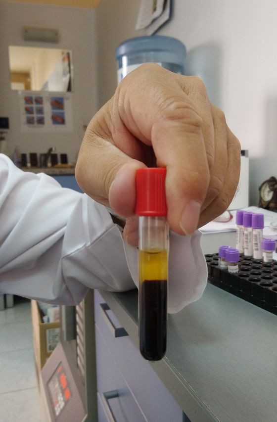
Serum is the fluid left after blood clots. Plasma is the fluid from blood that was kept from clotting. Most chemistry tests can be run on either serum or heparin plasma.1 The differences matter for a few analytes:
- Protein reads higher in plasma than serum, because fibrinogen is used up during clotting.4
- Potassium reads higher in serum, because cells release potassium during clotting. Merck notes potassium is best measured on heparin plasma separated right after collection.14
- Glucose is best kept in fluoride/oxalate (gray top) plasma when the sample cannot be separated quickly. Fluoride reads about 7 to 12% lower than promptly separated serum, because it lyses red cells and their water dilutes the sample.16
- EDTA (purple top) binds calcium and magnesium. Many enzymes need these ions, so EDTA plasma is not used for most chemistry.4
Analyzer types
In-clinic chemistry analyzers use one of two methods.1
- Wet chemistry (transmission or absorbance photometry): liquid reagents react with the sample and light passes through it. This is the reference method that reference values are based on.
- Dry chemistry (reflectance photometry): the sample is put on a slide or pad that holds dry reagent, and the analyzer reads light reflected from the color change. Merck notes these methods do not always compare well with the reference method and are best for simpler tests such as glucose and urea.
Whatever the analyzer, run control samples of known value at least daily for each analyte, in both normal and abnormal ranges. If the controls are out of tolerance, do not test patient samples.1 In-house results are best treated as an interim answer; Merck advises confirming them at a reference laboratory when needed.1
Renal analytes
- Urea (BUN) rises with dehydration, renal failure, a blocked urethra or ruptured bladder, and also with high dietary protein or catabolic states. It falls with liver failure and portosystemic shunts.1
- Creatinine rises with renal dysfunction, a blocked urethra and ruptured bladder. Animals with a large muscle mass run high-normal values; thin animals run low-normal.1
- SDMA (symmetric dimethylarginine) is cleared mainly by the kidneys. In one study of cats with chronic kidney disease, SDMA rose before creatinine in 17 of 21 cats, by a mean of 17 months.10
Hepatic analytes
- ALT (alanine aminotransferase) lives in liver cells and rises with hepatocellular damage in dogs and cats.1
- ALP (alkaline phosphatase) rises with steroids, biliary disease, bone growth and other causes. Its half-life is 72 hours in dogs but only 6 hours in cats. Cat values are much lower, so any increase in a cat is significant.1
- GGT (gamma-glutamyl transferase) rises with longer-term liver damage and is especially useful in horses and ruminants.1
- Bilirubin rises with hemolysis, liver dysfunction, biliary obstruction and fasting in horses. Yellow plasma is a cue to measure it, except in horses and cattle, whose plasma is normally yellow.1 Protect bilirubin samples from light.8
- Bile acids test liver function. They are more sensitive than bilirubin to liver impairment, and a portosystemic shunt causes a marked rise after a meal.1
Glucose, proteins and electrolytes
Glucose rises after carbohydrate meals, with stress or excitement (including handling and sampling), with glucocorticoids and with diabetes mellitus. It falls with insulin overdose and insulinoma.1 Stress is the most common cause of an isolated high glucose.6 Total protein rises with dehydration and chronic inflammation. Albumin rises with dehydration and falls with liver failure and protein loss. Globulin is calculated: total protein minus albumin.1 Sodium and potassium are the key emergency electrolytes; potassium rises with hypoadrenocorticism and severe renal failure, and falls with vomiting and diarrhea.1
| Analyte | Main organ | Common causes of increase | Sample trap |
|---|---|---|---|
| BUN (urea) | Kidney | Dehydration, renal failure, blocked urethra, high-protein diet | Also rises with diet and dehydration |
| Creatinine | Kidney | Renal dysfunction, blocked urethra, ruptured bladder | High-normal in muscular animals |
| ALT | Liver (dog, cat) | Hepatocellular damage | Decreases as the sample deteriorates |
| ALP | Liver, bone | Steroids, biliary disease, bone growth | EDTA inhibits many enzymes |
| GGT | Liver | Longer-term liver damage (horse, ruminant) | Use serum or heparin plasma |
| Bilirubin | Liver, red cells | Hemolysis, liver dysfunction, biliary obstruction | Protect from light; lipemia and hemolysis can raise it |
| Glucose | Pancreas, whole body | Stress, meals, steroids, diabetes | Falls if cells are not separated |
| Potassium | Kidney, adrenal | Hypoadrenocorticism, severe renal failure | Hemolysis, EDTA and serum raise it |
| Calcium | Parathyroid, bone | Dehydration, hyperparathyroidism, some cancers | EDTA and citrate lower it |
Sources: Merck Veterinary Manual, Clinical Biochemistry;1 eClinPath sample collection, interferences and bilirubin.458
The three interferences
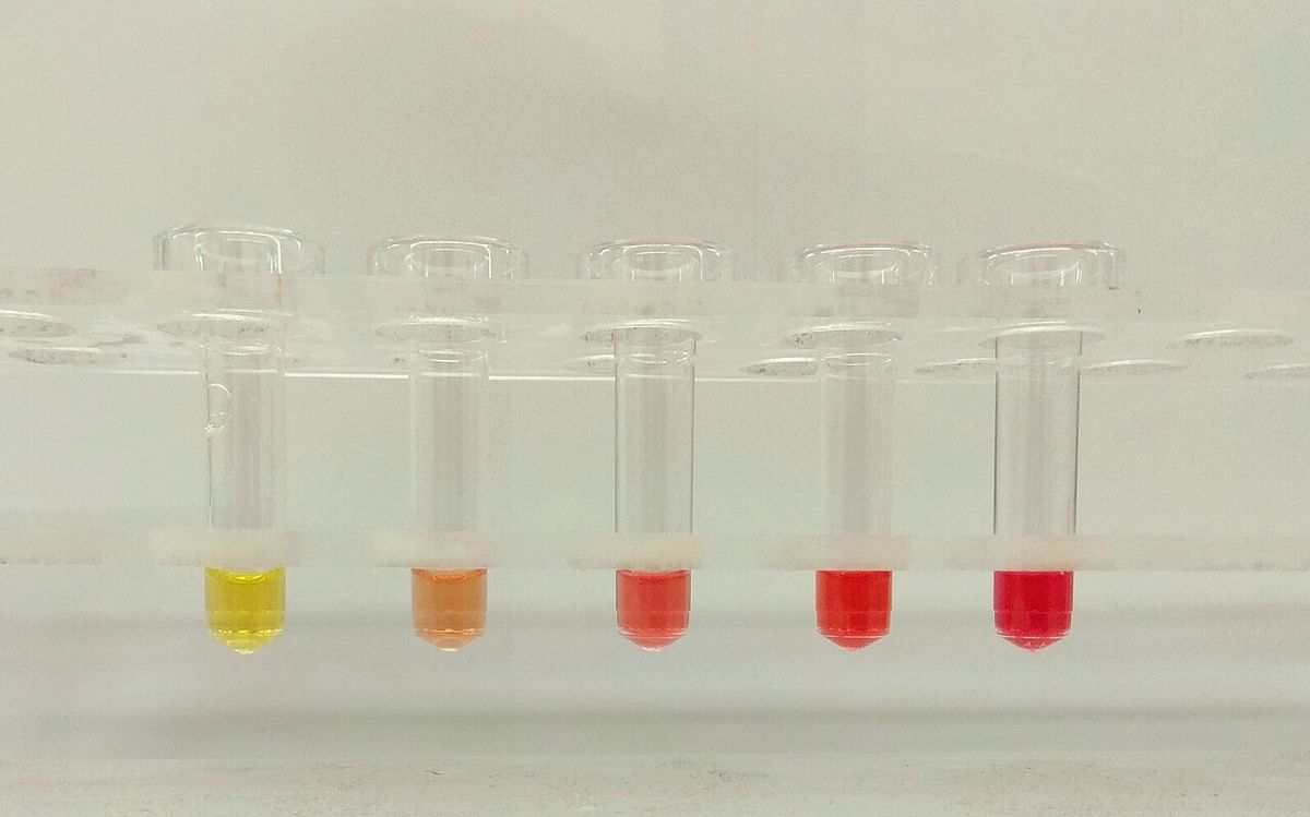
Hemolysis is the release of hemoglobin from broken red cells, seen as pink to red serum. In the tube it usually comes from poor venipuncture, lipemia, freezing whole blood, delayed separation or fluoride/oxalate tubes.5 Broken cells release potassium, AST, LDH and magnesium, falsely raising them. Potassium is most affected in species with high red cell potassium: horses, camelids, some cattle and Asian dog breeds such as the Akita and Shiba Inu.5
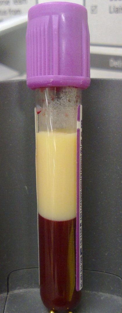
Lipemia is milky serum from triglycerides. It is usually a post-meal artifact, and a fast of at least 12 hours reduces it.5 Lipemia scatters light and can falsely raise some results, such as total bilirubin with some methods. It also displaces water, falsely lowering sodium and chloride. It makes red cells more fragile, so lipemic samples hemolyze more easily.5
Icterus is yellow plasma from high bilirubin. With some analyzers, bilirubin itself interferes with other tests.5 Remember that yellow plasma is normal in horses and cattle.1
Blood gas basics
A blood gas analyzer measures pH, partial pressure of carbon dioxide (pCO2) and partial pressure of oxygen (pO2), and calculates bicarbonate. Blood pH in health stays within about 7.35 to 7.45.13 pCO2 is controlled by breathing, so it shows the respiratory side; bicarbonate shows the metabolic side. Low pH is acidemia; high pH is alkalemia.913 Venous samples are fine for pH and pCO2, but pO2 is only reliable from an arterial sample.9
Procedure or technique
Preparing a serum or plasma chemistry sample
- Confirm the test list and any fasting instructions with the veterinarian. Fasting at least 12 hours limits lipemia.5
- Make sure no EDTA from a purple-top tube or needle carries into the chemistry tube. Even minor contamination changes potassium and calcium.4
- For heparin tubes, fill them more than half full to avoid artifacts such as falsely raised bilirubin.4
- For red-top tubes, let the blood clot for at least 30 minutes. Large animal blood clots slowly; loosen the clot from the tube wall with a wooden applicator stick.14
- Centrifuge. Merck gives 3,000 rpm for 10 minutes in a bucket centrifuge.1
- Pipette the serum or plasma into a fresh, labelled tube unless the gel tube gives a permanent separation. Serum separator gel does not stop storage artifacts if blood sits on it.14
- Look at the serum. Record hemolysis, lipemia or icterus on the request or in the record.
- Run controls, then the sample. If shipping, keep it cool on ice packs, without direct contact with the ice.4
Bile acid panel
- Collect a fasting sample, preferably after an overnight fast or just before the usual meal.7
- Feed a standard meal.
- Collect the post-prandial sample 2 hours after eating.7
- Label each tube with the time and "pre" or "post".
Blood gas sample
- Use a commercial heparinized blood gas syringe, or coat a syringe with sodium or lithium heparin and expel it. Keep heparin below 5% of the blood volume; too much heparin changes the results.9
- Collect from a peripheral artery (for oxygenation) or a vein.9
- Expel any air bubble and cap the syringe at once. Air falsely lowers pCO2; 10% air bubbles will affect results.9
- Run the sample promptly. Blood gas is a point-of-care test and should not be mailed to a laboratory.9
Flowchart
Species differences
- Cats: prone to stress hyperglycemia from handling, so in-hospital glucose curves may be questionable.11 ALP is normally much lower than in dogs, so any rise matters.1
- Dogs: ALP in the thousands is usually linked to raised steroid levels.1 Akitas and Shiba Inus have high red cell potassium, so hemolysis raises their potassium.5
- Horses and ruminants: GGT and GDH (glutamate dehydrogenase) are the more useful liver enzymes. Plasma is normally yellow. Blood clots slowly, so leave time before spinning.14 Horses have high red cell potassium, so leaving serum on the cells can raise potassium.4
| Analyte | Dog | Cat | Horse |
|---|---|---|---|
| Urea nitrogen (mg/dL) | 8 to 28 | 19 to 34 | 11 to 27 |
| Creatinine (mg/dL) | 0.5 to 1.7 | 0.9 to 2.2 | Use lab range |
| Glucose (mg/dL) | 76 to 119 | 60 to 120 | 62 to 134 |
| Total protein (g/dL) | 5.4 to 7.5 | 6.0 to 7.9 | 5.6 to 7.6 |
| Sodium (mEq/L) | 142 to 152 | 146 to 156 | 128 to 142 |
| Potassium (mEq/L) | 3.9 to 5.1 | 3.7 to 6.1 | 2.9 to 4.6 |
| Blood pH | 7.31 to 7.42 | 7.24 to 7.40 | 7.32 to 7.44 |
| pCO2 (mmHg) | 29 to 42 | 29 to 42 | 36 to 46 |
Sources: Merck Veterinary Manual, Serum Biochemical Analysis Reference Ranges and Blood-Gas Analysis Reference Ranges.23 Ranges vary between laboratories and analyzers; always use the range printed by your lab.
Calculation
Worked example: globulin and SI units. A dog's panel shows total protein 7.2 g/dL and albumin 2.8 g/dL. Find the globulin, and give total protein in g/L.
Globulin = total protein minus albumin.1
7.2 g/dL minus 2.8 g/dL = 4.4 g/dL globulin.
To convert g/dL to g/L, multiply by 10.1 7.2 g/dL × 10 (dL/L) = 72 g/L; the dL units cancel and g/L remain.
Report the calculated value and let the veterinarian interpret it against the laboratory's own range.
Safety, legal and client education
- Treat every blood sample as potentially infectious. Wear gloves, keep the centrifuge lid closed until the rotor stops and dispose of needles as sharps.
- The veterinarian in charge of the practice laboratory is responsible for all results issued, which is why quality control records must be kept.1
- Tell clients when to withhold food before a panel or bile acid test, and explain that a meal can make serum milky and affect results.57
- For owners of diabetic cats, explain that the stress of a clinic visit can raise glucose, which is why home monitoring or fructosamine may be used.11
Common mistakes and exam traps
- Leaving serum on the clot. Glucose falls, and potassium can rise. Separate promptly; a gel tube does not prevent this.4
- Using EDTA for chemistry. High potassium with very low calcium suggests EDTA contamination.4
- Calling yellow horse plasma icteric. It is normal in horses and cattle.1
- Treating a high glucose in a stressed cat as diabetes. Stress hyperglycemia must be excluded first.11
- Leaving an air bubble in a blood gas syringe. It falsely lowers pCO2.9
- Running patients when controls fail. Merck says no patient samples should be tested.1
Memory aids
HLI, "the three that lie" (memory aid): Hemolysis, Lipemia, Icterus are the three common interferences; look for pink, milky or yellow serum before every run.
PURPLE PULLS CALCIUM (memory aid): EDTA in the purple top binds calcium, so contamination drops calcium and raises potassium.
PRE, MEAL, PLUS TWO (memory aid): bile acid panel order: a fasting sample, a standard meal, then a sample 2 hours later.
BUBBLES BLOW OFF CO2 (memory aid): air in a blood gas syringe falsely lowers pCO2, so expel bubbles and cap at once.
CATS SPIKE (memory aid): cats spike glucose with handling stress, usually below 288 mg/dL and gone within a few hours.
Quick review
- Serum is from clotted blood; plasma is from anticoagulated blood.
- Heparin plasma is the preferred anticoagulant sample for chemistry.
- Let red tops clot at least 30 minutes, then spin and separate promptly.
- Unseparated cells lower glucose; fluoride/oxalate preserves it.
- EDTA raises potassium and lowers calcium.
- Hemolysis falsely raises potassium, AST and LDH.
- Lipemia scatters light and lowers sodium and chloride; fast 12 hours.
- Wet chemistry is the reference method; dry slides use reflectance.
- Run controls at least daily; do not run patients if they fail.
- SDMA can rise before creatinine in kidney disease.
- Bile acids: fasting sample, meal, then 2-hour sample.
- Blood gas: heparin syringe, no air, run at once; pO2 needs arterial blood.